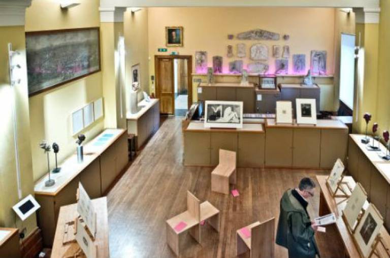
Using museum artwork to teach cardiac anatomy
George Richards (UCL Public and Cultural Engagement) shares what happened when students learned about the heart through two watercolours painted by a UCL professor of surgery and artist.

24 February 2016
As part of the UCL Art Museum’s drive to foster experimental teaching, we held a series of drop-in sessions on how to use objects and artfacts to teach last May.
These clinics were organised to illustrate the possibility of matching artworks from our collection with courses beyond the traditional scope of object-based learning.
We made a concerted attempt to attract lecturers from scientific and mathematical backgrounds, and were delighted to find that a number of lecturers found it useful.
Discovering a link between art and surgery
The Institute of Cardiovascular Science was one such department, and we are indebted to Dr. Ann Walker for her advocacy from the very outset.
Dr. Walker is the Graduate Tutor of the Institute, and attended the drop-in session with several of her colleagues to explore the possibility of including museum objects within their MSc module in Cardiovascular Science.
They recognised potential in two watercolours by artist and Professor of Surgery at UCL, Sir Charles Bell.
So as to further explain the history of the Bell drawings and their potential use in teaching, we also invited Dr. Chiara Ambrosio, a longstanding collaborator of the UCL Art Museum, to attend the clinic. Chiara uses the works to teach her module on the History and Philosophy of Science. She knows them intimately, and her enthusiasm for them is infectious. So much so, the Institute committed to basing a tutorial around them shortly afterwards.
Using art and specimens together to make discoveries
Dr Thomas Kador (Teaching Fellow, UCL Museums and Collections) had the idea of including additional UCL resources within this planning. He introduced Ann to Subhadra Das - the curator of the UCL Pathology Collections at the Royal Free Hospital - and together they identified a specimen that related to the tutorial - a fluid-preserved heart. The heart was couriered to the Art Museum shortly before the lecture commenced.
After a brief introduction, Chiara began by introducing the Bell watercolours, and explaining the surgeon’s link to UCL, as well as his contributions to anatomy and the nervous system.
Dr. Andrew Cook (Senior Lecturer) then invited his students to consider what Bell’s paintings and the specimen has in common.
The students soon recognised peculiarities in the representation of the phrenic nerves, as well as mitral stenosis in the specimen.
The uniting factor was therefore ablation therapy (one of the treatments for mitral stenosis; damage to the phrenic nerves can also be a complication caused by such therapy), and Dr. Cook explained this procedure in detail. Dr. Riyaz Patal - senior lecturer and consultant cardiologist at UCL - was also at hand to discuss atrial fibrillation.
Getting students engagement by using a new perspective
The change in the environment of study had brought about more engagement and participation from students.
On the back of this, there is now talk of developing another lecture next year around a drawing of the internal thoracic artery by progressive surgeon and fellow UCL alumnus, Joseph Lister.
This example serves to demonstrate just what can be accomplished when the strength and breadth of UCL expertise and resources are pooled together.
UCL Art Museum is part of UCL’s Public and Cultural Engagement Department.
 Close
Close

