XClose

UCL Institute of Ophthalmology

Home
Menu

What are Bestrophinopathies?

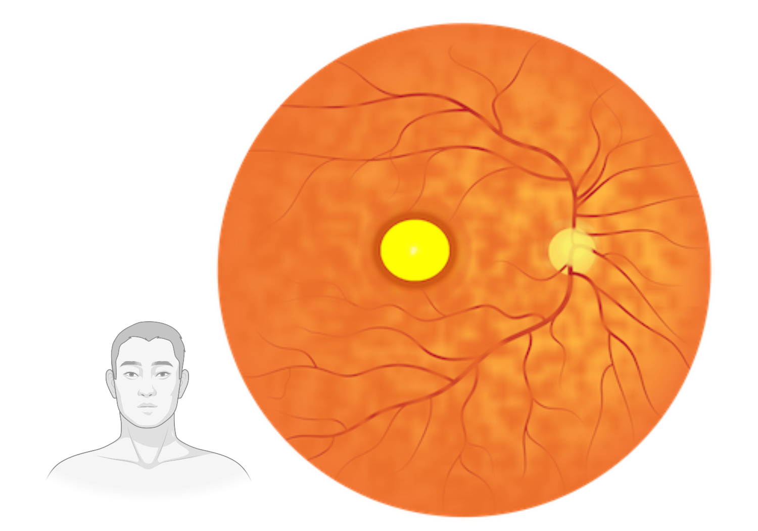
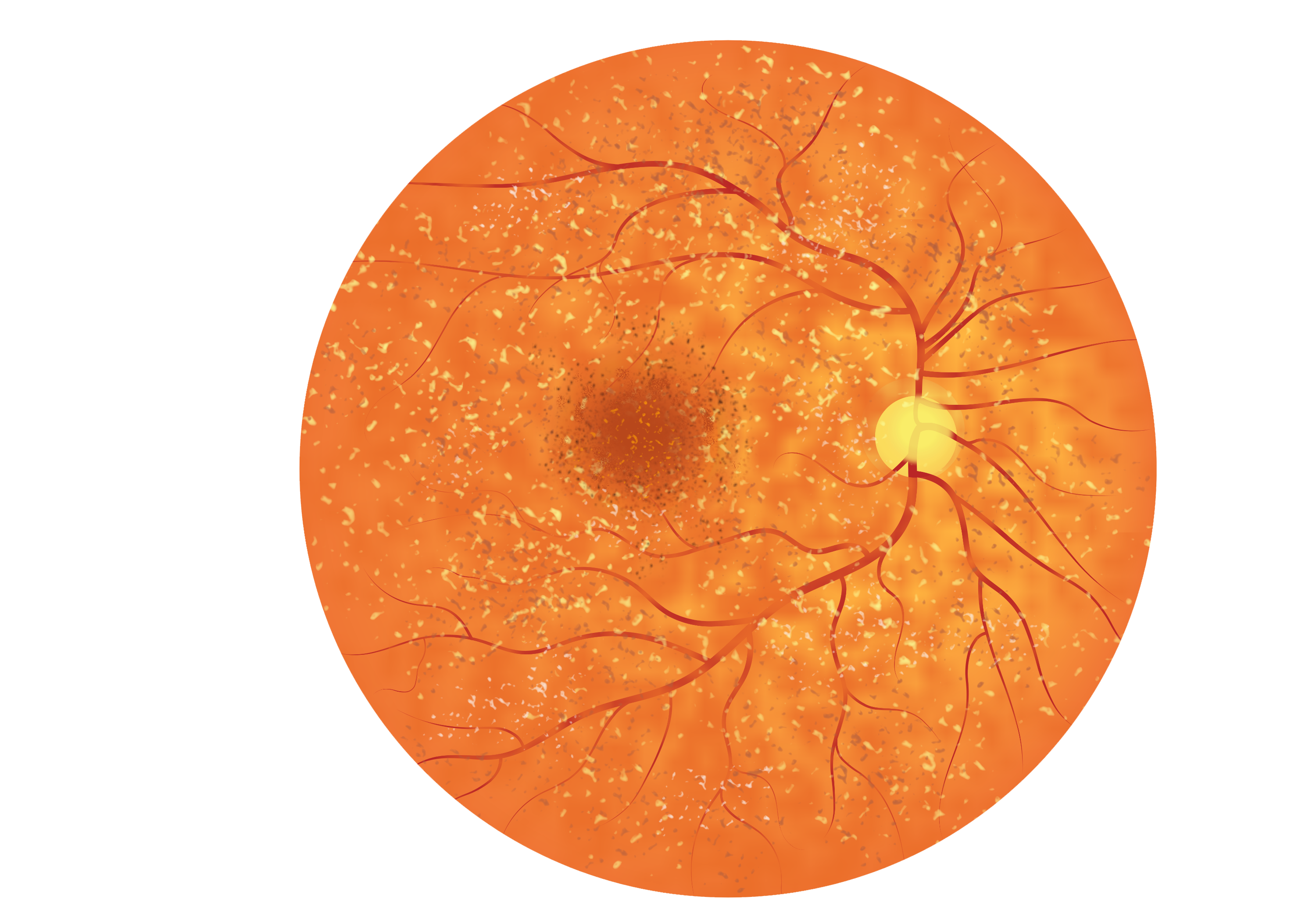
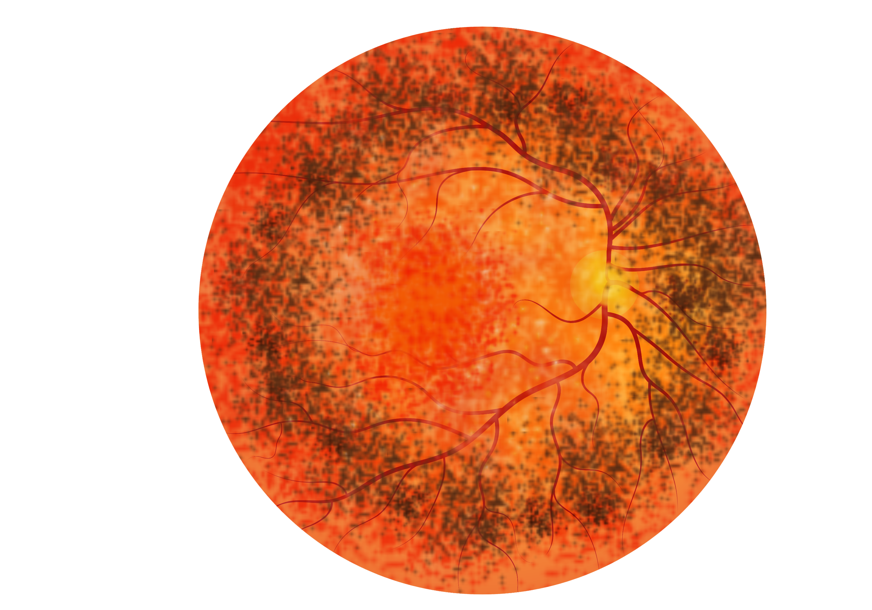
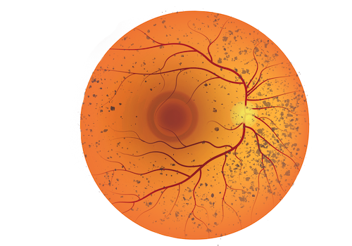
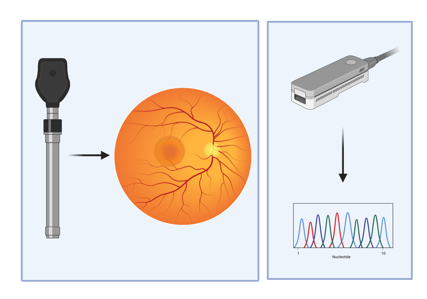
Bestrophinopathies are a group of clinically distinct diseases causing retinal degeneration and vision loss in both eyes. Bestrophinopathies are caused by mutations in a single gene, known as the Bestrophin-1 (BEST1) gene. Find out about the individual diseases by clicking the links below.
Fundus image of a retina suffering from Best disease. Visible 'egg-yolk' formation.

Best Disease

Find out more about Juvenile Best Disease

Fundus image of a retina suffering from AVMD. Visible 'egg-yolk' formation.

Adult Vitelliform Macular Degeneration

Find out more about the adult form of Best’s Disease

Fundus image of a retina suffering from Autosomal Recessive Bestrophinopathy. Fundus appears speckled, with irregularities of the RPE cells and the presence of yellow/white dots or flecks scattered across the retina.

Autosomal Recessive Bestrophinopathy (ARB)

Find out more about Autosomal Recessive Bestrophinopathy (ARB)

Schematic of the fundus image of a retina suffering from ADVIRC. A highly pigmented circular band present in the outer regions (periphery) of the retina, with a distinct border between the highly pigmented retina and normal retina.

Autosomal Dominant Vitreoretinochoroidopathy (ADVIRC)

Fine out more about Autosomal Dominant Vitreoretinochoroidopathy (ADVIRC)

Fundus image of a retina suffering from Retinitis Pigmentosa. Peripheral retina pigment deposits can be seen and a swelling macular

Retinitis Pigmentosa (RP)

Find out about Retinitis Pigmentosa (RP)

Left: Ophthalmoscope producing a fundus image. Right: Sequencer producing a sequencing trace.

How is a Bestrophinopathy Diagnosed?

There are different tests and read-outs which diagnose and distinguish the bestrophinopathies