 Close
Close

Translation imaging: Visible biomaterials
13 March 2024
Making implanted meshes visible
A translation imaging collaboration between CABI (Stephen Patrick, Mark Lythgoe, Daniel Stuckey) and CMI (Steve Halligan, Sam Parker) to make implanted meshes visible. This bench to bedside approach is published in Advanced Science, and presents the first method distinguishing surgical meshes used in abdominal hernia surgery from normal bodily tissue.
Prototypes of surgical meshes that can be seen clearly on CT scans have been developed by Dr Stephen Patrick and colleagues for the first time, promising to increase the speed at which problems can be diagnosed and resolved.
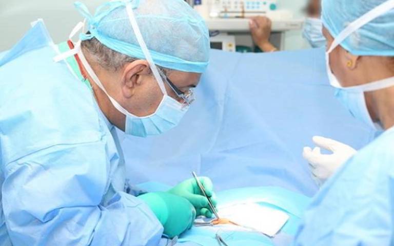
Hernia happens when an internal part of the body pushes through the surrounding muscle and tissue, creating a bulge. Around a quarter of men will experience a hernia in their lifetime, making hernia surgery the most commonly performed operation in the world.
As these surgical meshes are commonly made from biological material, they bind with the surrounding tissue to reinforce the area – but this also means the mesh becomes difficult to distinguish from normal tissue, something that creates real problems when these patients need re-operation because surgeons cannot determine what has been done before.
In this study, researchers from UCL were challenged by surgeons at UCLH to come up with a solution to identify implanted surgical meshes.
They repurposed an old type of chemistry, normally used to bind radioactive iodine isotopes to proteins such as antibodies for the purposes of detecting diseases. By swapping the radioactive iodine for a stable, non-radioactive isotope, they were able to engineer them into protein-based surgical meshes so that they would show up more clearly on CT scans, which are used commonly for planning surgery.
Dr Stephen Patrick, senior author of the study from UCL Division of Medicine, said: “When colleagues at UCLH first posed the problem of surgical meshes being difficult to distinguish from normal tissue, our thoughts quickly turned to how we could label the material to make it visible to CT. In the end, we repurposed an old type of chemistry to do something new and to enhance X-ray absorption. The beauty of the approach is that it is also easy and cheap, so I’m confident that it could make a difference to patients in the near future.”
The authors are exploring how to incorporate the technique into commercial meshes with producers in order to get them into the clinic where they will be able to address issues of visibility.
Professor Steve Halligan, an author of the study and a radiologist from UCL Division of Medicine, said: “The results of our study are very exciting. Ventral hernias are increasingly common and while most patients won’t experience any issues after repair, when problems do occur they can be very tricky to resolve. We’re already talking to manufacturers with a view to getting the technology into the clinic and start making a difference for patients as soon as possible.”
Cheltenham Science Festival
Dr Stephen Patrick has now extended this work to Imaging Microparticles, which will be unveiled at Cheltenham Science Festival Sat 8 Jun 2024 alongside a paper and press release.
Title: What are microplastics, how do we find them and should we worry?
They float around in our bottled water, our deepest oceans, the clouds above us, and even the human blood stream. Tiny specks of eroded plastic from that long-forgotten Christmas toy, food wrappers and clothing are everywhere. Using the latest imaging technology, Mark Lythgoe, Stephen Patrick and Mark Miodownik track down these elusive particles on stage, investigating the mysteries of where they come from, how they get into our bodies, and how we can limit our consumption.
Links
- Dr Stephen Patrick’s academic profile
- Professor Steve Halligan's academic profile
- Professor Mark Lythgoe Profile
- Miss Lady Barrios Silva's academic profile
- UCL Centre for Advanced Biomedical Imaging
- UCL Division of Medicine
- UCL Faculty of Medical Sciences
- Cheltenham Science Festival Sat 8 Jun 2024