Location: Basement (Room B09), Queen Square House.
The electron microscopy unit provides expert services for the preparation of semithin resin sections, ultra-thin sections and electron microscopy grids, and teased nerve fibres for diagnostic purposes.
Electron microscopy examinations of the grids are performed by our staff. The EM is located in the Camelia Botnar laboratories at Great Ormond Street hospitals.
Please be advised that the Electron Microscopy facility at the Queen Square Institute of Neurology has been decommissioned in 2021. We currently offer ultrastructural examinations only for diagnostic samples.
Contacts:
Enquiries for diagnostic and research services
Angela Richard-Londt, Head Biomedical scientist,
Email: angela.richard-londt@nhs.net
Enquiries for diagnostic services
Dr Michael Groves, Biomedical Scientist
Email: m.groves@ucl.ac.uk / michael.groves@nhs.net
Examples of specimen preparations in our EM Unit
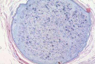
Peripheral nerve; semithin resin section, MBA-BF stain

Peripheral nerve: teased fibre preparation

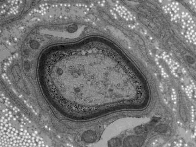
Peripheral nerve: Electron microscopy; widely spaced myelin.
Price List
| Transmission Electron Microscopy sample preparation charges. Prices for Electron microscopy examination on request. | |
| All prices excl. VAT | |
| Standard machine processing into resin, per run | £55 |
| Hand processing into resin, per run | £115 |
| Preparation of semithin section, each block | £38 |
| Preparation of set of stained grids, each block | £85 |
 Close
Close


