 Close
Close

DOT-HUB

Research
Read about our main areas of research and current projects below.
- The ANIMATE project

The ANIMATE project — Wearable neuroimaging technologies for the study infant motor development
Newborn infants vulnerable to brain injury and often go on to develop cerebral palsy. Cerebral palsy is a group of permanent movement disorders that can severely limit the control of the muscles, and can have a devastating impact on quality of life. Cerebral palsy is also the most common form of childhood disability in Europe and every year, approximately 1,800 children in the UK are diagnosed with the condition. The early diagnosis of cerebral palsy is critical. While there is no cure for the condition, there are a number of treatments that can improve an infant's long-term motor ability. At present, the majority of infants with cerebral palsy are not diagnosed until 1 or 2 years-of-age. By this point it is likely too late for treatment to have the best possible impact. If infants with abnormal motor development could be identified early, these treatments would have the greatest chance of success. We think brain imaging can help understand and diagnose cerebral palsy. However, no current functional imaging available with the necessary precision, resolution, patient comfort, and motion tolerance.
The ANIMATE project is funded by the EPSRC and aims to develop a new wearable functional brain imaging technology to investigate the emergence of cerebral palsy in infants at the cot-side. The project is based at DOT-HUB, UCL and neoLAB, but depends upon critical collaborations with Prof. John Suckling (Cambridge University), Dr. Tom Arichi (Kings College London), Dr. Betty Hutchon (The Royal Free), Prof. Frances Cowan (Imperial College London) and industrial partners Gowerlabs.Current research on this project involves many members of DOT-HUB, including Dr. Hubin Zhao, Elisabetta Maria Frijia and Liam Collins-Jones
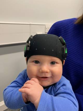
- LUMO applications
- LUMO applications

In collaboration with Gowerlabs, we are undertaking a series of proof-of-principal studies using the LUMO high-density diffuse optical tomography system. These studies involve a range of experiments in a range of populations to test the performance of this new generation of technologies.
This work is being led by Dr. Ernesto Vidal Rosas and Elisabetta Maria Frijia.

- Infant anatomical head modelling
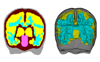
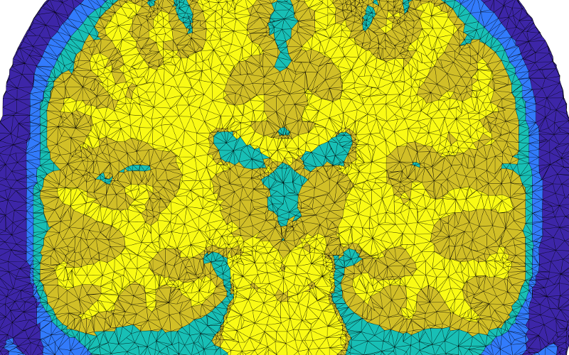
Infant anatomical head modelling
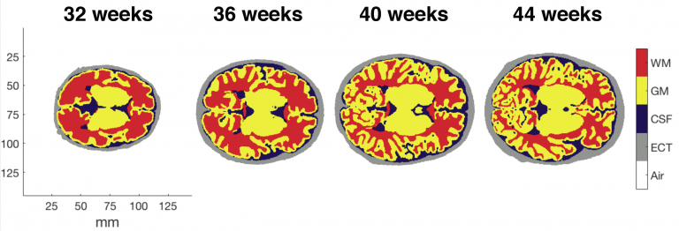
The infant head undergoes rapid structural changes on a timescale of weeks. Diffuse optical tomography (DOT) requires a model of photon transport from sources to detectors through a subject's head - and this requires structural information. A structural prior is a model of the head wherein tissues with different optical properties are delineated. Ideally, a structural prior would be produced using a subject's own MRI scan. However, necessitating an MRI scan for each subject undermines the benefits of DOT, notably its portability and its tolerance of motion that allows the infant brain to be studied in the awake state. At DOT-HUB we are developing anatomical models of the infant head using state-of-the-art MRI data from the Developing Human Connectome Project (dHCP) that can be used when modelling photon propagation within the head. At present we are developing a database of structural for infants aged 28-44 weeks post-conception and quantifying the effect on DOT image quality of using a structural prior from the database as opposed to a subject-specific structural prior.
- Wearable HD-DOT development
- Wearable HD-DOT development

As part of the ANIMATE project, we have developed a neonatal-specific wearable high-density DOT module. It exploits flexible electronics to produce ultra-low profile, lightweight sensors that can be directly interconnected to form imaging arrays. By combining various modules together via in-built board-to-board connector, the system can be used to create wide range of ultra-lightweight, flexible HD-DOT imaging arrays. This will incorporate hundreds of emitters and detectors of near-infrared light to safely image the whole cortex of infant brain.
YouTube Widget Placeholderhttps://www.youtube.com/watch?v=I5lUQUru4Go
