 Close
Close

We offer access to a wide range of scientific equipment, notably the microscopes of the Research Department of Surgical Biotechnology at the Royal Free Campus in Hampstead.
Light Microscopes
- High resolution Leica TCS SP8 Confocal Microscope (upright)
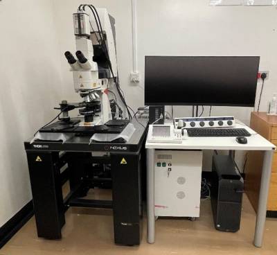
Provides a multicolour 3D imaging platform with optimal photon efficiency and high-speed LIGHTNING, providing ultimate sensitivity and precision. The system is fully automated and well suited to imaging a wide range of live and fixed multi-labelled cells and tissues, cell-seeded non-transparent scaffolds, implants or in well plates as well as small animals.
Key features
- 4 detectors (2 GaAsP HyD and 2 PMT) and a motorized xyz-stage.
- Objectives: 10x 0.4, 20x 0.75, 40x 1.3 (oil), 63x 1.4 (oil).
- Laser lines: 5 laser lines (405, 448, 488, 552 and 638nm).
- Incubator chamber for live cell imaging.
Location
9th Floor, Department of Surgical Biotechnology, Royal Free Campus.
(Training and ClusterMarket login required.)
- Olympus fluorescent microscope BX63 (upright)
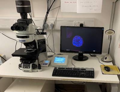
The Olympus wide field fluorescent upright microscope BX63 is easy to use for imaging both dry and subversive samples.
Key features
- Variety of objectives for dry imaging (x4, x10, x20, x 40) and submersive (x10, x20, x40).
- Filter Cubes are for (DAPI, FITC, TRIC, CY5, CY7, CY8) as well as bright field imaging.
- The excitation is with Cool-LED PE300.
Location
9th Floor, Department of Surgical Biotechnology, Royal Free Campus.
(Training and ClusterMarket login required.)
Electron Microscopes
- Zeiss Field Emission Scanning Electron Microscope, FESEM Sigma 300VP
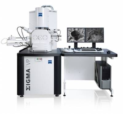
The latest model of field emission scanning microscope with high-end imaging and advanced analytical techniques. Surfaces, nanostructures and chemical composition of materials, cells, microorganisms and tissues can be imaged in both 2D and 3D with the highest quality by choosing from a variety of detector options.
Key features
- Field emission, GEMINI column, variable pressure (VP) technology.
- InLens SE Detector: for high resolution in column topographical imaging.
- ETSE Detector: for high vacuum topographical imaging at longer working distance.
- AsB Detector: compositional and crystallographic orientation imaging.
- VPSE-G4 Detector: Fourth generation Variable Pressure SE detector.
- aSTEM Detector: Annular STEM for transmission imaging.
- SmartEDX: energy dispersive X-ray spectroscopy (EDS) analysis system with multi-point spectra, line-scan, mapping, quantification drift correction and reporting.
- 3D Surface modelling: provide a 3D reconstructed topographical view and measure the texture of the sample surface.
Location
Analytical Imaging Suite, Low-Ground, Department of Surgical Biotechnology, Royal Free Campus.
(Training and ClusterMarket login required.)
- Zeiss Scanning Electron Microscope, Zeiss MA10
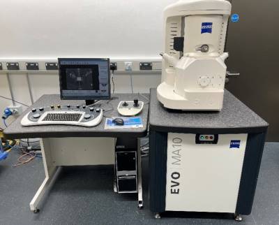
The Zeiss EVO MA10 is an easy-to-use benchtop SEM which provides superior performance with good resolution and high throughput.
Location
Analytical Imaging Suite, Low-Ground, Department of Surgical Biotechnology, Royal Free Campus.
(Training and ClusterMarket login required.)
- Transmission Electron Microscope Philips CM12, 100keV
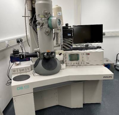
The Philips CM12 is a general purpose analytical transmission transmission electron microscope (TEM). It is used to visualize the structure, chemical composition, and arrangement of atoms in thin, extraordinary small specimens.
Key features
- Thermionic electron source.
- Selectable beam acceleration voltage, 20 kV to 120kV.
- Magnification range 31x – 660,000x.
- Transmission (TEM), selected area diffraction (SAD), bright field and dark field operating modes.
- X-Ray Energy Dispersive Spectrometer (EDS) for elemental analysis.
- CCD camera.
Location
Analytical Imaging Suite, Low-Ground, Department of Surgical Biotechnology, Royal Free Campus.
(Training and ClusterMarket login required.)
Stanmore Campus
- Research facilities
- GLP and research tissue culture facilities: For culture expansion including primary culture of various human and animal cells.
- Molecular biology laboratory: Including Fluidigm, Ion Proton and Ion Torrent systems.
- Microscopy suites: FM, SEM, TEM and CM (at Eastman Dental Institute (EDI)).
- Atomic Force Microscope (EDI).
- General Biochemistry Laboratory (plate readers, spectroscopy suite, BondMax Autostainer). Mechanobiology laboratories: Including custom built cell culture force and tensioning monitors, Instron testing machine, DMA testing kit (BME and EDI).
- Finite Element Analysis software.
- Image Analysis Software.
- Histology Laboratories: Including Exact Cutting and grinding machinery for hard grade resin histology.
- Gait analysis laboratory (ACDS).
- Development and characterisation of advanced drug delivery systems: Rotary evaporator, ultra-sonicator, freeze-dryer and fluorescence microscope.
- Laser and low intensity ultrasound systems for treatments for cancer.
- Facilities for in vivo tumour model development: For the assessment of advanced drug delivery systems, pharmacokinetics, toxicology.
- Orthopaedic implant design and manufacture: Dedicated workshop (including orthopaedic implant testing facilities, mills and lathes for orthopaedic implant design and manufacture.
- Dedicated IT hubs supported by UCL.
- Electronic library access and specialised Orthopaedics library (on the Royal National Orthopaedic Hospital (RNOH) site).
- Clinical research facilities.
- Protein materials manufacture and characterisation laboratories.
- Walk in fridge / freezer rooms / humid 37oC room / -70 oC freezers and liquid nitrogen storage.
- Dual beam UV-VIS spectrometer 1nm bandwidth (lambda 25, Perkin Elmer).
- Fluorescent plate reader extended for NIR fluorescence (BMG LABTECH CLARIOstar,).
- Flat-bed imaging system including NIR filter pair (G:BOX CHEMI HR1.4, Syngene).
- Small animal endoscopy system (Karl Storz).
- Range of red and NIR lasers with a range of different laser delivery fibre systems. (Various)
- Cat I tissue culture suite with 3 hoods.
- Expertise
- GLP and tissue culture: For various human and animal cells (including, but not exclusively, fibroblasts, tenocytes, myoblasts, osteoblasts, neuronal cells, primary pulmonary artery smooth muscle cells (inner and outer) and mesenchymal stem cells).
- Molecular biology techniques: Including RT-PCR, Southern blotting, Western blotting, Northern blotting, plasmid transfection of human and animal cells. Whole/targeted genome, whole exome and whole transcriptome sequencing.
- Microscopy: Fluorescence (FM), Scanning Electron (SEM) and Transmission Electron (TEM) and confocal (CM).
- Atomic Force Microscopy.
- Biochemistry: including Flow Cytometry, High Performance Liquid Chromatography (HPLC), ELISA, Mass Spectroscopy, immunocytochemistry.
- In Vitro Mechanobiology.
- Finite Element Analysis.
- Image Analysis: For the analysis of histological specimens.
- Histology: Hard grade resin histological techniques for hard tissue sectioning with metal implants. Wax histology and immunohistochemistry.
- Gait Analysis: Human and animal.
- Development and characterisation of advanced drug delivery systems.
- Laser and low intensity ultrasound treatments: For cancer, cardiovascular disease and infection prevention, as well as the development of formulations that are responsive to these forms of energy.
- In vivo tumour model development.
- Orthopaedic implant design and manufacture: at The Centre for Materials.
Royal Free Campus
- Research facilities
- Advanced biomaterials laboratories for manufacturing nanomaterials, polymers, ceramics, hydrogel and 3D scaffolds for tissue engineering and regenerative medicine
- Manufacturing and implant design workshop including plasma surface modifier, electrospinning/electrospraying machine, nitric oxide analyser, impedance spectrum analyser, thromboelastography (TEG®) Hemostasis Analyzer System, and 11 3D printers with 3D image reconstruction and design software
- Nano-spray coating technology including ultrasound vapour atomisation, spin coating, sputter coating, plasma modification
- Precision Matlab® potentiostats
- Environmental SEM and confocal microscopy
- Dedicated material analysis laboratory with viscometer/rheometer, solid phase peptide synthesis, UV-Spectrophotometer and luminescence spectrometer, drop shape analyser, mechanical testing (Instron), laser cutter, dynamic light scattering and zeta potential analysis
- Nanocomposite laboratory with reactor ready bioreactor, Fourier Transform Infrared Spectrometer and IRTON-infrared microscope. Also access to custom built graft extruder at Stevenage
- Facilities in histology and immunohistochemistry including microtome, tissue embedding, freeze dryer, and automated tissue processing
- Dedicated tissue culture laboratories with nine Cat II biosafety cabinets for cell culture including cell lines and primary culture of various human, animal and stem cell lines (fibroblasts from skin, colon, breast, cervix, osteoblasts, myoblasts, neuronal culture, iPSCs, mesenchymal stem cells)
- Molecular biology facilities including Real-time RT-PCR and ChemiDoc gel imaging system
- General biochemistry with plate readers and spectrophotometers
- Facilities and expertise for the development and characterization of advanced drug delivery systems, including rotary evaporator, and ultra-sonicator
- Facilities in laser and low intensity ultrasound experimental treatments for cancer, as well as the development of formulations, which are responsive to these forms of energy
- Facilities in in vivo tumour model development, assessing advanced drug delivery systems, and pharmacokinetics
- Facilities in in vivo models of regenerative surgery, assessing implantation of tissue engineered constructs
- Dedicated small animal operating theatre for evaluating in vivo studies with doppler ultrasound
- Protein materials manufacture and characterization laboratories
- -80 freezers and liquid nitrogen storage facilities
- AFM and the Acoustic-piezoelectric system.
- Expertise
- Histology and immunohistochemistry
- in vivo models of regenerative surgery
- in vivo tumour model development
- 3D culture from cell lines and primary cells
- Optical therapy diagnostics and photodynamic therapy - laser and low intensity ultrasound experimental treatments for cancer and the development of light activated antibody drug conjugates
- Cancer epigenetics and genomics
- Statistical analysis including statistical genomics
- Stem cell biology and immunology in cell therapy
- Experimental cancer therapeutics, including gold nanoparticles for the imaging of colorectal cancer using X-ray diffraction and fluorescence
- Quantum Dots for dual imaging and delivery of therapy to colorectal cancer.
- Cryopreservation and transplantation for clinical applications
- Bioactive glasses
- Bio-manufacturing of tissue scaffolds and nanomedicines by 3D printing, electrospinning, flow-processing in combination with self-assembly of biomaterials;
- Design and fabrication of artificial organs, tissue engineering scaffolds, implantable biosensors and devices
- Surface modification of biomaterials - electrostatic coatings, self-assembled monolayers, thin films, polyelectrolytes, and surfactants, plus characterisation, e.g. Wettability/AFM/ESEM/XPS.
- Chemical modification of microelectrode biomaterials for electrostimulation (e.g. sensory & human nervous system applications)
- Synthetic biology and biomimicry using molecular hydrogel nanocomposites for 3D scaffolds for cardiac and neural tissue engineering.
- Cardiovascular bioengineering – Shape memory and alloys for stents, silicone elastomers, nanocoatings and nanocomposites.
- Neurological biomaterials – Neuro- and immunomodulation, chemical-biological modification, electrostimulation, IR induced delivery of therapeutic agents.
- Manufacture of colloids/nanodispersions and nanomaterials, tailored biological modification, toxicology and drug delivery
- Expertise in integrative analysis of genomic, histological, and imaging data.
Bloomsbury Campus
- Research facilities
- Dual beam UV-VIS spectrometer 1nm bandwidth (lambda 25, Perkin Elmer).
- Fluorescent plate reader extended for NIR fluorescence (BMG LABTECH CLARIOstar,).
- Flat-bed imaging system including NIR filter pair (G:BOX CHEMI HR1.4, Syngene).
- Small animal endoscopy system (Karl Storz).
- Range of red and NIR lasers with a range of different laser delivery fibre systems. (Various)
- Facilities and expertise in whole genome, whole exome and whole transcriptome sequencing using Ion Proton
- Facilities and expertise in targeted genome and transcriptome sequencing using Fluidigm and Ion Torrent
- Facilities and expertise in histology and immunohistochemistry using the BondMax Autostainer
- Facilities and expertise for the development and characterization of advanced drug delivery systems, including rotary evaporator, ultra-sonicator, freeze-dryer and fluorescence microscope.
- Facilities and expertise in laser and low intensity ultrasound treatments for cancer, as well as the development of formulations that are responsive to these forms of energy.
- Cat I tissue culture suite with 3 hoods.
- Facilities and expertise inin vivo tumour model development, assessing advanced drug delivery systems, pharmacokinetics, toxicology.
- Facilities and expertise in laser treatments for cancer, cardiovascular disease and infection prevention.
- Expertise
- Expertise in cancer epigenetics and genomics
- Expertise in high density DNA methylation/SNP array
- Epigenetic biomarker discovery, validation and targeted high throughput assay development
- Expertise in next generating sequencing and bioinformatic analysis of Bis-Seq/Methyl-seq, Chip-Seq, RNA-Seq and Exome data.
- Expertise in integration of large scale epigenomic, genomic and transcriptomic data and functional candidate validation
- Expertise in bioinformatics7. Expertise in single cell transcriptomics and whole genome sequencing.
- Expertise software engineering and computer programming
- Expertise in statistical analysis including statistical genomics
- Expertise in integrative analysis of big data (genomics, histology and imaging)
- Expertise in digital image processing and analysis from histology and radiological scanners (including 3D reconstruction from 2D slices)
- Expertise in mathematical modelling including non-linear systems
- Expertise in 3D cell culture systems.
- Photodynamic therapy, emphasis on its application in gasterenterology and head and neck cancers.
- Development of light activated antibody drug conjugates.
- Biomarker identification and validation of tissue and fluidic markers.
- Expertise in target validation for drug development.
- Expertise in ELISA development and validation including MSD.