XClose

UCL Division of Medicine

Home
Menu

Computational Medicine

The Centre for Computational Medicine is a multidisciplinary, cross-faculty research centre that draws together research in physics, mathematics, computer science and medicine. It combines UCL’s formidable strengths in mathematical and computational modelling of disease to create a unique and world-leading centre. We support an interdisciplinary, dynamic, inclusive environment that enables disruptive and creative thinking.

Our work

We draw on many disciplines to enable our research, including physics, mathematics, biology, chemistry, computer science, engineering and statistics. Many of our projects lie at the intersection of biomedical imaging, mathematical modelling and machine learning.

Biomedical Imaging

We use biomedical imaging to guide computational analysis, alongside developing novel, cutting-edge imaging techniques with a focus on translation and providing patient benefit. Ongoing project areas include:

Magnetic Resonance Imaging (MRI) of Cancer

We are developing quantitative MRI techniques to characterise the tumour microenvironment and predict drug delivery and treatment response. This includes techniques such as diffusion MRI modelling of tumour microstructure to non-invasively measure cell size and arterial spin labelling to measure vascular perfusion. (Recent paper)

Imaging Transplanted Organs as Model Systems

We are passionate about understanding, validating and translating new imaging approaches, and are developing methods for trialling new technologies in transplanted human organs that are maintained by mechanical perfusion. This provides a novel platform to investigate real tumours in situ and how they interact with imaging biophysics (collaboration with Royal Free Hospital and WEISS).

Optical Imaging

We develop new optical imaging solutions such as high-resolution episcopic microscopy (HREM) and light-sheet microscopy. This allows us to create large-scale digital tissue models (‘digital twins’) that can be used for mathematical modelling and for better understanding biomedical imaging techniques.

HiP-CT

Mapping human organs at multiple scales using synchrotron radiation. (Recent paper)

HiP-TC

Opt brain vessels

Brain lectin

Mathematical Modelling

We use mathematical modelling to explore and quantify the relationship between function and form.

Blood Flow and Solid Mechanics in Tumours

REANIMATE is our framework for combining three-dimensional microscopy of tumours and in vivo imaging, to model blood flow, and interstitial fluid dynamics to better understand and optimise drug delivery to tumours. The interaction between fluid mechanics and solid mechanics is an active area of investigation, particularly in relation to metastatic growth in the spine (collaboration with Leeds University). (Recent paper)

Blood Vessel Network Simulation

We have developed several platforms for modelling normal blood vessel networks, using biophysical principles such as Murray’s Law, alongside models of aberrant growth such as angiogenesis in tumours.

OncoEng

An EPSRC-funded project in collaboration with the University of Leeds, aiming to predict fracture risk in spinal metastases. (Project website: OncoEng).

Retinal modelling

We are creating detailed, large-scale models of the human retina, using ophthalmology image data, to better understand the link between vascular delivery and metabolic demand (collaboration with Moorfields Eye Hospital and Institute of Ophthalmology). Understanding the cause of loss of vascular perfusion in diabetic retinopathy is a specific motivation and its impact on vision. 

Liver Perfusion

Using a range of MRI approaches, we are developing large-scale models of liver vascular perfusion, particularly during the growth of tumours, and investigating the delivery of therapeutic nanoparticles (collaboration with Royal Free Hospital and UCL Chemistry).

REANIMATE (3D microscopy of tumours)

Synthetic vessels

Retina vessels

Machine learning / Artificial Intelligence

Physics informed deep learning

Our imaging and modelling work combines physics-informed deep learning, in which mathematical models are used to train generative learning algorithms. For example, working with colleagues at Moorfields Eye Hospital, we are developing deep generative modelling approaches to segment and classify images from the retina.

Supervised learning

We have developed a large imaging database of vascular imaging data with manual labels, which forms the basis of our tUbe-Net framework for segmenting blood vessel networks with minimal manual labelling (more on GitHub).

Retina CycleGAN

Blood vessel structure (tUbe-Net)

Our experts 

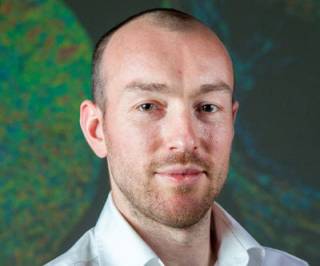
Simon Walker-Samuel portrait

Prof. Simon Walker-Samuel

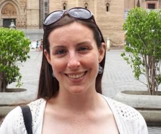
Prof. Rebecca Shipley

Natalie Holroyd

Dr Natalie Holroyd

Basic silhouette in a circle, in light grey

Dr Andrew Guy

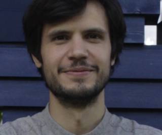
Simao Laranjeira

Dr Simão Laranjeira Gomes

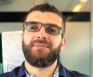
Hani Cheikh Sleiman

Dr Hani Sleiman

Basic silhouette in a circle, in light grey

Dr Maxime Berg


PhD Students
  • Emmeline Brown 
  • Grant Lauder 
  • Lucie Gourmet 
  • Emre Doganay 
  • Hannah Coleman 
  • Shawn (Li) Zhongwang
  • James (Jie) Lam
Affiliated Members 
  • Claire Walsh 
  • Dr Paul Sweeney

Selected Publications

  1. d'Esposito A, Sweeney P, Shipley R, Walker-Samuel S, et al. Computational fluid dynamics with imaging of cleared tissue and of in vivo perfusion predicts drug uptake and treatment responses in tumours. Nature Biomedical Engineering, 2018, 2:773–787.
  2. Walsh CL, Walker-Samuel S, Brown E, Holroyd N, et al. Imaging intact human organs with local resolution of cellular structures using hierarchical phase-contrast tomography. Nat Methods. 2021 Dec;18(12):1532-1541.
  3. Gourmet LE, Walker-Samuel S. The role of physics in multiomics and cancer evolution. Front Oncol. 2023 Mar 17;13:1068053.
  4. Holroyd NA, Walsh C, Gourmet L, Walker-Samuel S. Quantitative Image Processing for Three-Dimensional Episcopic Images of Biological Structures: Current State and Future Directions. Biomedicines. 2023 Mar 15;11(3):909.
  5. West H,  Sweeney P, Walker-Samuel S,  Shipley RJ, et al. A mathematical investigation into the uptake kinetics of nanoparticles in vitro. PLoS One. 2021 Jul 22;16(7):e0254208.
  6. Berg M, Holroyd N, Walsh C, Walker-Samuel S, Shipley R, et al. Challenges and opportunities of integrating imaging and mathematical modelling to interrogate biological processes. Int J Biochem Cell Biol. 2022 May;146:106195.
  7. Roberts TA, Hyare H, Walker-Samuel S, et al. Noninvasive diffusion magnetic resonance imaging of brain tumour cell size for the early detection of therapeutic response. Sci Rep. 2020 Jun 8;10(1):9223. 
  1. Breen-Norris JO, Walsh C, Walker-Samuel S, et al. Measuring diffusion exchange across the cell membrane with DEXSY (Diffusion Exchange Spectroscopy). Magn Reson Med. 2020 Sep;84(3):1543-1551.
  2. Jafree DJ, Walsh CL, Walker-Samuel S, et al. Spatiotemporal dynamics and heterogeneity of renal lymphatics in mammalian development and cystic kidney disease. Elife. 2019 Dec 6;8:e48183. 
  3. Sweeney PW, Walker-Samuel S, Shipley RJ, et al. Modelling the transport of fluid through heterogeneous, whole tumours in silico. PLoS Comput Biol. 2019 Jun 21;15(6):e1006751. 
  4. Sweeney PW, Walker-Samuel S, Shipley RJ. Insights into cerebral haemodynamics and oxygenation utilising in vivo mural cell imaging and mathematical modelling. Scientific Reports, 2018; 8:1373.
  5. Zhu Y, Bradley D, Walker-Samuel S, et al. Non-invasive imaging of disrupted protein homeostasis induced by proteasome inhibitor treatment using chemical exchange saturation transfer MRI. Scientific Reports, 2018; 8: 15068
  6. Gonçalves MR, Johnson SP, Walker-Samuel S. The effect of imatinib therapy on tumour cycling hypoxia, tissue oxygenation and vascular reactivity. Wellcome Open Res 2017, 2:38
  7. Gonçalves MR, Peter Johnson S, Walker-Samuel S, et al. Decomposition of spontaneous fluctuations in tumour oxygenation using BOLD MRI and independent component analysis. Br J Cancer. 2016 Jun 14;114(12):e13

Funding and Partnerships

Logo for Cancer Research UK. A large mosaic C of blue and pink circles plus dark blue text.

Wellcome Trust logo

Logo for the ESPRC (Pioneering research and skills)

The logo for the URKI Medical Research Council. A quadrilateral, with 'UKRI' over navy on the left, and two teal portions on the right.

A logo featuring a styled RT containing a rose. Text reads: "Rosetrees Trust. Supporting the best in medical research"

Logo for the Chan Zuckerberg Initiative

Interested in joining us?

Researchers: We regularly have funding for postdoctoral positions and welcome prospective applications from talented postdoctoral candidates seeking to obtain their own funding. To discuss potential opportunities, please contact Prof. Simon Walker-Samuel (simon.walkersamuel@ucl.ac.uk) or Prof. Rebecca Shipley (rebecca.shipley@ucl.ac.uk). Please include a CV with your email.

Students: If you have a good academic record and would like to discuss opportunities in our Centre, please contact Prof. Simon Walker-Samuel (simon.walkersamuel@ucl.ac.uk) or Prof Rebecca Shipley (rebecca.shipley@ucl.ac.uk), including a CV with your email.

Contact details

Email: compmed@ucl.ac.uk

Twitter: @swalkersamuel

Postal Address:
Centre for Computational Medicine
Division of Medicine
Gower St
LONDON
WC1E 6BT

Visiting Address:
Centre for Computational Medicine
Division of Medicine
Ground floor, Rayne Building
5 University Street
LONDON, WC1E 6JF