Close
Close

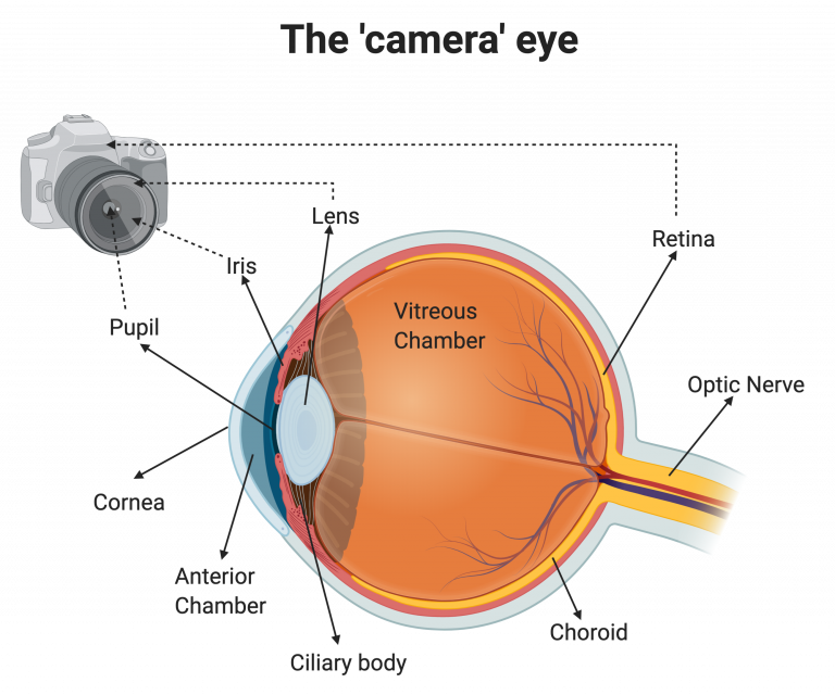
The human eye, despite all its complex nature, can be described and compared to a modern photo camera
The human eye consists of several components which work in tandem with the aim to efficiently capture light and turn it into an image in the brain. Light enters the pupil of our eye, much like light enters the front of a camera. Our pupils act as a natural aperture, increasing or decreasing in diameter to capture more or less light from our surroundings as needed. This change in pupil size is governed by the muscles of the iris, the coloured part of the eye in which the pupil is the central opening. The iris can be compared to the shutter of a camera, as it controls how much light is able to enter through the pupil.
Before light enters through our pupil, it passes through the transparent part of the eye known as the cornea. The cornea is a transparent structure with no blood vessels, which acts as the primary focusing power of the eye, bending light so that it converges on the retina to form an image. Once light is refracted by the cornea it travels through the pupil into an anterior chamber full of aqueous humour, a liquid providing essential nutrients to the eye. Light then meets another transparent structure behind the pupil known as the lens. The shape of the lens can be altered by a group of muscles, collectively known as the ciliary body, changing the precise point of light focus on the retina. Like a camera, the lens helps us to focus light when looking at close objects, or those at a distance. The focused light then travels through a large vitreous chamber filled with a transparent gel-like protective substance, the vitreous humour.
Light then reaches the light sensitive part of the eye, the retina. The retina is a highly complicated structure which can be best thought of as the film of the camera. The retina is responsible for taking light and developing it into electrical information, which can be sent to the brain via the optic nerve to enable us to see an image correctly. The retina is supported by the choroid, a rich blood vessel network that supplies the retina with the oxygen and nutrients required to keep the retina working. The retina and choroid are physically separated by a barrier of cells, known as the retinal pigment epithelium (RPE)