Lab. Head: Professor Roger Lemon
Premotor-motor cortex interactions during visually-guided grasp
Understanding grasp-related LFPs in primate motor cortex
Cortical mechanisms subserving visually-guided grasp in humans
This project investigates how the brain implements motor commands for grasping movements. The secure grasp of an object requires appropriate hand shaping and precise force scaling, two processes controlled by a specific cortical ‘grasping circuit’ constituted of several anatomically
distinct but interconnected cortical areas. We have developed a special paired-pulse
paradigm for use with non-invasive transcranial magnetic stimulation (TMS) in human volunteers. We are using this paradigm to investigate cortico-cortical interactions between one area of this grasping circuit and its inputs to M1. This approach is being combined with several motor tasks which allow us to study cortico-cortical interactions
during different contexts of hand shape or force generation. We are particularly interested to determine which cortical areas are involved and when activity in these pathways is able
to influence hand shape prior to grasp.
In collaboration with the Wellcome Trust Centre for Neuroimaging (WTCN), we also use concurrent TMS-fMRI to study causal cortical activation patterns in motor areas during different grasp contexts, using an MRI-compatible graspable device.
Neurophysiological mechanisms of tool use by monkeys and humans
Mirror neurons
The discovery of mirror neurons in the premotor cortex of macaque monkeys generated some major new concepts in cognitive and motor neuroscience. A large number of non-invasive investigations have indicated that a similar 'mirror neuron system' exists in humans. Mirror neurons have been implicated in several different roles, including understanding the actions and intentions of others, prediction and imitation. In addition, it has been suggested that disturbance of this system could contribute to autism. Moreover, activity of mirror neurons during action observation could be used to train brain machine interfaces operated by paralysed individuals. However, our knowledge of the distribution of mirror neurons in the motor and premotor cortex and of their input and output connectivity is still rudimentary.
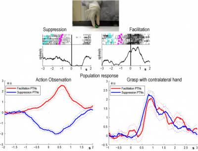
We recently showed that identified pyramidal tract neurons (PTNs) in area F5 of the premotor cortex could show mirror-like properties, which means that mirror activity influences sensorimotor function in the spinal cord. Interestingly, about half of these PTNs, although active when the monkey grasped, showed complete suppression of their discharge when monkeys observed an experimenter grasping an object. We are currently following up these observations to see if these suppression mirror neurons could act to suppress the monkey’s own movements while it watches the actions of others.

Reference Kraskov A, Dancause N, Quallo MM, Shepherd S, Lemon RN (2009) Corticospinal neurons in macaque ventral premotor cortex with mirror properties: a potential mechanism for action suppression? Neuron 64:922-930. PMC free text
Neurophysiology and neuroanatomy of small-diameter corticospinal axons
Small fibres are ubiquitous in the mammalian corticospinal tract. Our understanding of the functions of these small fibres, which far outnumber large ones, is still rudimentary. These fibres may be very important in clinical conditions such as spinal cord injury because not only are they much more numerous than large axons, but they are much less susceptible to trauma. Together with our UCL collaborator, Mitch Glickstein, here we are looking at the spectrum of axon diameters in the corticospinal and corticopontine systems and their cortical areas of origin. We are combining this with a study of the physiological response properties of small-diameter corticospinal fibres to intracortical and non-invasive stimuli.
 Close
Close

