Image Archive

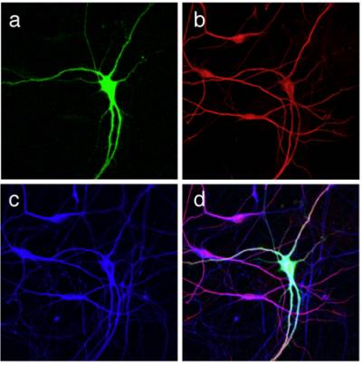
Figure 2 and 3. Differentiation of mouse ES cells into MNs. An optimised protocol allows the differentiation of primary MNs from a mouse ES cell line. MN specification is determined by the addition of retinoic acid and sonic hedgehog, whilst MN maturation is achieved in the presence of neurotrophins. ES cell-derived MNs are characterized by an HB9-driven GFP staining (green) and are positive for the bIII isoform of tubulin (red), and for the dendritic marker MAP2 (blue).
 Close
Close

