 Close
Close

New technology helps to identify throat cancer more precisely during surgery
27 April 2023
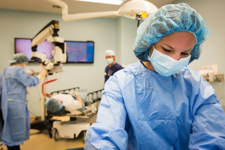
New research from WEISS reveals that a new type of endoscope has been developed to detect how polarised light interacts with tissue. This has the potential to be integrated into endoscopes to provide a new method of detecting disease, including cancer, during surgery, allowing better decisions to be made during the procedure and helping to save lives.
Published this month in Nature Biomedical Engineering, the study was a collaboration between scientists from WEISS, Imperial College London, Zhejiang Lab, China, and Northwick Park Hospital, London. The team set out to solve the issue that current endoscopes and techniques used during throat cancer surgery need to be more sensitive and can lead to unsatisfactory rates of false negatives. The group developed a new method for detecting diseased tissue to solve this problem, termed ‘surgical polarimetric endoscopy’ (SPE).
The interaction of polarised light with tissue can reveal properties of the tissue that are not visible to the naked eye. For instance, tissues with a clear oriented structure, such as collagen fibres, present a different signal to tissues with disordered structures. Since the progression of a cancer often involves the breakdown of organised tissue structures into a disorganised tumour, imaging of the polarisation has been suggested as a new method to detect this disease. The benefits of using light are significant; it is non-harmful and can be adapted onto current surgical devices such as cameras and endoscopes, saving the NHS money in the long run. In addition, this new technique records these signals quickly and efficiently during surgery.
Dan Stoyanov, Professor of Robot Vision, WEISS, said: “Visualization is crucial in keyhole surgery. Discovering new ways to see more information than is available to the naked eye can greatly enhance surgical care. This study is an example of an exciting new development.”
Daniel Elson, Professor of Surgical Imaging and Biophotonics, Hamlyn Centre for Robotic Surgery and Department of Surgery and Cancer, Imperial College London, states: “The use of commercially available endoscopes adapted to acquire polarisation information has enabled us to acquire the data during head and neck procedures but could in principle be expanded to other keyhole surgery applications. The real-time acquisition and display of data is also very important for the surgeon since it makes it easier for them to use and test the endoscope.”
The researchers and clinicians involved in the study were able to design, build and test the endoscope in a human study during head and neck procedures. This shows the potential of the method for intraoperative decision-making, helping surgeons to see changes in the tissue not visible to the naked eye. In addition, they reviewed how the polarisation properties changed between normal and cancerous laryngeal tissue and created videos showing the data being acquired at video rate.
Dr Ji Qi, Research Center for Humanoid Sensing, Zhejiang Lab, Hangzhou, China, said: “This study demonstrates the proposed endoscopy with polarisation image contrast is an effective complement to routine endoscopy, and the combination of the proposed and routine endoscopy can enhance intraoperative laryngeal cancer detection. This may therefore open up new opportunities for better intraoperative guidance and improved surgical care of head and neck tumours.”
The clinical feasibility of this method has now been established, allowing a more extensive evaluation on further specimens and other procedures and diseases. Mr Taranjit Tatla, Consultant Head and Neck Surgeon, Northwick Park Hospital, said: “Further clinical application and validation of this novel low-cost tool requires continued collaborative, cross-organisational and inter-disciplinary teamwork for digital data collection and correlation to gold standard histopathology. Integrated into a modular multi-modal augmented vision platform, designed to support both objective endoscopic diagnostics and precision micro and robotic surgery across multiple organ systems and diseases, its value is especially to be realised through the incorporation of machine learning and artificial intelligence software.”