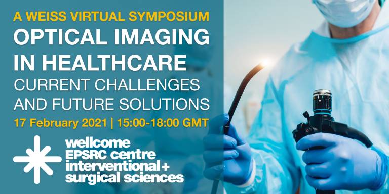
Optical Imaging in Healthcare: Current Challenges and Future Solutions
24 February 2021
On 17 February 2021 we hosted our 6th WEISS virtual symposium, this time on the topic of optical imaging in healthcare.

This symposium, chaired by Dale Waterhouse, brought together world-leading imaging researchers with track records of clinical translation to help showcase the biggest successes of novel imaging technologies as well as summarising the key outstanding barriers to clinical implementation and discussing future strategies to accelerate translation.
Brian Pogue (Dartmouth College) began the symposium with his talk titled ‘Biomedical Optics: The single largest technology sector in medicine’. He provided an overview of biomedical optics, describing the enormous variation in design and economics of clinical optical devices, ranging from otoscopy and ophthalmology to robotic surgery.
This was followed by a presentation from Sylvain Gioux (Intuitive Surgical Inc.) on the current progress and challenges for spatial frequency domain imaging for surgical guidance.
Next, Dimitris Gorpas (Technical University of Munich) discussed the challenges and prospects for near infrared fluorescence image-guided interventions, using the example of intra-operative tumour-specific fluorescent imaging in ovarian cancer by folate receptor-a targeting.
Laura Marcu (University of California, Davis) spoke about intra-procedural fluorescence lifetime imaging (iFLIM), focusing on the iFLIM technology that exploits the unique fluorescence signatures of endogenous biomolecules to provide a map of tissue biochemical features and metabolic state.
Thomas Wang (University of Michigan) discussed early cancer detection in the digestive tract. Certain cell surface targets are over overexpressed in cancerous cells, so Thomas and his group have been developing a strategy to image these targets using peptide imaging agents.
Elizabeth Hillman’s (Columbia University) talk covered high-speed 3D microscopy for in-vivo, in-situ, instant histology. Her lab has been developing a technology called SCAPE (swept confocally aligned planar excitation) microscopy which is a form of light sheet microscopy. This has the ability to perform motionless ultra-fast 3D acquisition at high sensitivity and low phototoxicity.
Following this, Nick Stone (University of Exeter) gave his talk ‘Raman spectroscopy: more than a real-time, point-of-care, spectroscopic measure of pathology?’
George Gordon (University of Nottingham) discussed holographic imaging through optical fibres. For the purposes of his talk, he defined holographic imaging as the use of coherent light for illumination and also using coherent detection of the light coming back.
Frédéric Leblond (Polytechnique Montreal) spoke about clinical translation challenges in Raman spectroscopy, specifically for surgical applications. Raman spectroscopy is a technique used to determine vibrational modes of molecules, providing a structural ‘fingerprint’ by which molecules can be identified in a non-invasive way.
The symposium had a fantastic turn out of 115 people from 16 different countries. We look forward to hosting more symposia in the near future – make sure to keep checking the Events pages on our website for details!
 Close
Close

