Close
Close

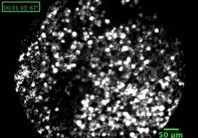
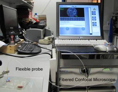
A Fibered Confocal Microscope works as a conventional Confocal Microscope except that the objective lens has been replaced with a flexible probe made up of tens of thousands of optical fibres for in vivo observations of the tissues in real-time. The probe is placed in contact with the tissue to scan its surface with a laser beam that may have a wavelength of either 488nm or 660nm. There is a large range of probes for observations at depths varying from the tissue surface to 100μm with optical thicknesses of about 10μm. The microscope can work in fluorescence or in reflectance mode. For fluorescence imaging, the tissue has to be stained with an appropriate fluorophore. The acquired images have a field of view of approximately 500μm x 500μm with a resolution of 3μm. Morphological and functional imaging can be performed.
The Fibered Confocal Microscope is used clinically for GI cancer research. The probe is passed via the working channel of a widefield endoscope to interrogate the tissue usually stained with fluorescein. It is also used for small animal studies, for example, for peripheral nerve imaging, observation of neurons with cellular resolution, or angiogenesis imaging. In CABI, the Fibered Confocal Microscope has been mainly used for the study of the dynamic stained cells with a 10μm diameter.
See the collaborations page for information on access to this system