 Close
Close

Electrical Impedance Tomography is a recently developed imaging technique, with which images of the internal impedance of the subject can be rapidly collected with rings of external ECG-type electrode

It is fast, inexpensive, portable and sensitive to physiological changes which affect electrical impedance properties. For about two decades, satisfactory images have been obtained of changes over time related to gastric emptying and ventilation and cardiac output in the thorax.
The work of our group at UCL has been to pioneer EIT for imaging brain function. It could be used to image in acute stroke or epileptic seizures, when its portability and low cost would give it unique practical advantages over existing methods such as fMRI. It could also provide images of fast neural activity in the brain over milliseconds which would constitute a revolutionary advance in neuroscience technology.
Innovations in hardware and image reconstruction algorithms enable accurate images to be collected in tanks and in experimental animals with electrodes on the brain; the next challenge is to see if recent technical improvements allow us to collect clinically useful images in human subjects with scalp electrodes.
Key advances
- Development of idea that EIT could provide tomographic images of fast electrical activity in the brain and so provide a uniquely new method to test theories in computational neuroscience (Holder, 1987)
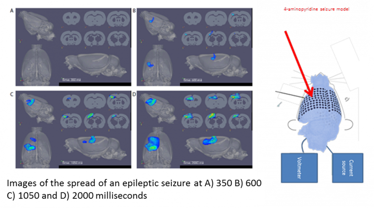
Demonstration that EIT with the Sheffield Mark 1 EIT system could produce reproducible EIT images of stroke (Holder, 1992), epileptic seizures, functional activity (Holder et al, 1996, Oh et al, 2011) and the phenomenon of spreading depression which is thought to underlie migraine (Boone et al, 1994) in anaesthetised experimental animals (rats or rabbits) with a ring of electrodes on exposed brain
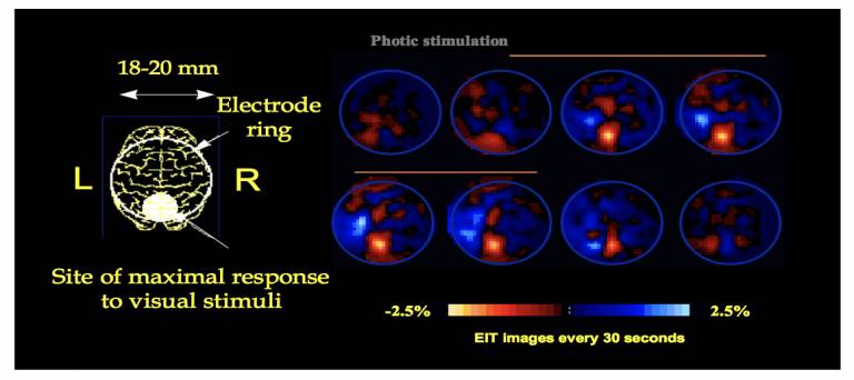 Fig 1: fMRI-like EIT images during visual evoked activity in the anaesthetised rabbit with cortical electrodes (Holder et al, 1997).
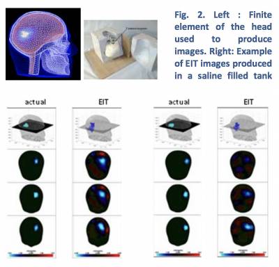
- Development of image reconstruction software able to produce accurate EIT images of brain function in 3D with scalp electrodes, using anatomically realistic Finite Element Models of the brain (Bagshaw et al, 2003). Refinements include anatomically realistic Finite Element Model meshes (Vonach et al, 2012, Aristovich et al, 2014), adjustment for anisotropy (Abascal et al, 2008), improved regularization methods (Aristovich et al, 2014), and novel methods for imaging at a single moment in time in acute stroke based of recording with multiple frequencies (Malone et al, 2013, Malone et al, 2014).
Development of electronic hardware specially tailored for EIT imaging in acute stroke (Yerworth et al, 2003; McEwan et al, 2006, Oh et al, 2007), epileptic seizures (Yerworth et al 2002) and fast neural activity in the brain (Avery et al, 2017).
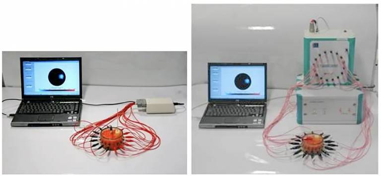
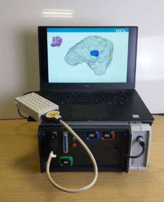
Fig 3: Examples of EIT systems developed at UCL. Left : UCH Mk2.5 (single channel record and current drive, multiplexed to 16 or 32 electrodes, switches serially, ~ 3 images per second (McEwan et al, 2006) Middle: KHU Mark 1 (16 parallel record single drive, ~ 1 image per second; Oh et al, 2007) Right : UCL ScouseTom human EIT system (2019)
- First clinical studies in humans of EIT in stroke (Romsauerova et al, 2006), epileptic seizures (Fabrizi et al, 2006) and blood flow related changes over seconds in normal cortical evoked activity (Tidswell et al, 2001).
- A recent breakthrough has been the production of the first tomographic images of fast neural activity during cortical evoked activity or epilepsy in the anaesthetized rat using epidural electrode mats (Aristovich et al, 2015, Hannan et al, 2020).
Use of fast neural EIT with a nerve cuff for imaging activity in the fascicles inside peripheral nerves. There is a new field in medicine – “Electroceuticals”. Autonomic nerves are stimulated electrically with the aim of treating a broad range of endocrine, autoimmune and other disease. However, current approaches stimulate entire autonomic nerves such as the vagus nerve in the neck; this produces unwanted side effects. With Glaxo Smith Kline and Google Health (Verily) we have developed a flexible nerve cuff that enables imaging of activation of individual pathways inside nerves i.e. spatially-selective vagus nerve stimulation (sVNS). This offers a promising solution to mitigate the off-target effects associated with traditional VNS and enhance therapeutic outcomes. However, this requires the knowledge of the fascicular organization at the level of cuff placement to inform selectivity of only the desired target organ or function.
Our group has recently determined the organisation of cardiac, pulmonary and recurrent laryngeal activity in the cervical vagus nerve at the level of cuff placement. We imaged function over milliseconds with fast EIT and selective stimulation, and found consistent spatially separated regions within the nerve correlating with the three fascicular groups of interest, suggesting organotopy. This was independently verified with structural imaging by tracing anatomical connections from the end organ with micro-computed tomography and the development of an anatomical map of the vagus nerve. This confirmed organotopic organization of the cardiac, pulmonary and recurrent laryngeal fascicles and function. These findings pave the way for improved outcomes in VNS and extension of this technique to treat other disorders. (Thompson et al, 2023)
Work in progress is mapping the cardiac efferent and afferent projections, specifically. This would serve as a precise method for addressing chronic heart failure (HF) by specifically targeting efferent cardiac fibers to reduce heart rate and override the sympathetic overactivation.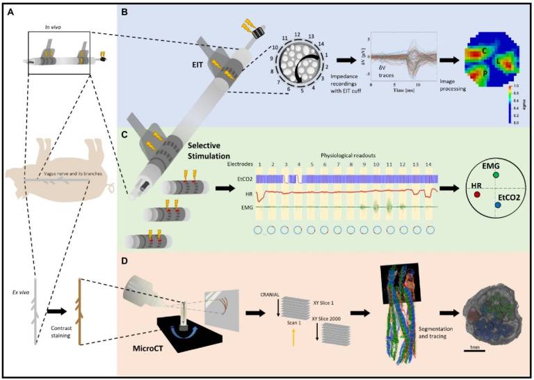
Experimental design for pig cervical vagus nerve imaging with Electrical Impedance Tomography (EIT), selective stimulation (SS), and micro-computed tomography (microCT). (A) In vivo experiment in pigs with EIT and SS cuffs placed around the left vagus nerve (N = 4) followed by dissection of the nerve from cervical level to below the pulmonary branches, including the cardiac and laryngeal branches, for ex vivo microCT. One additional bipolar cuff is placed around the recurrent laryngeal (RL) branch to evoke action potentials. (B) Identification of the areas responsible for cardiac (C), pulmonary (P), and laryngeal (L) functions in the cervical vagus nerve with EIT. The color scale is arbitrary units (Z score of relative change in the modulus of the impedance). (C) Selective stimulation through individual electrode pairs applied sequentially around the circumference of the cuff (pairs 1–14) with resulting physiological changes, such as heart rate (HR, red, cardiac), electromyography (EMG, green, recurrent laryngeal) and end-tidal carbon dioxide (EtCO2, blue, pulmonary), to determine cross-sectional location of the fascicular groups responsible for the respective functions. (D) MicroCT scanning of the full dissected vagus nerve followed by segmentation and tracing from the point of organ-specific branching up to the cervical level of cuff placement to identify the location of organ-specific fiber-containing fascicles (cardiac, red; laryngeal, green; and pulmonary, blue).
Current projects
Projects in progress in the group are :
- Imaging fast neural circuits in the brain in health and diseases with EIT with intracerebral depth probes
- Non-invasive imaging of human brain function over milliseconds with magnetic detection EIT with MEG or time-of-flight CHIRP EIT.
- Selective vagal nerve stimulation using fast neural EIT in heart failure.