 Close
Close

World Cancer Day 2022
4 February 2022
This World Cancer Day we're championing advances in cancer research and treatment made by our staff, students and partners.

New x-ray imaging scanner will assist surgeons while performing breast cancer tumour removal surgery
Through the £4.8m Prosperity Partnership, UCL and Nikon joined forces to develop new techniques to gain more information from X-rays, improve disease detection and industrial testing.
One of the advances brought about by this partnership was the co-development by UCL Medical Physics and Biomedical Engineering researchers and Nikon of a new x-ray imaging scanner that will assist surgeons while performing breast cancer tumour removal surgery.
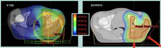
This new approach to x-ray imaging allows surgeons to assess extracted tissue intraoperatively or during the initial surgery, giving 2.5 times better detection of diseased tissue in the margins than with standard imaging. Researchers used X-Ray Phase Contrast Imaging (XPCI), developing a scanner which provides surgeons with a full 3D image of the extracted tissue lump, known as a wide local excision (WLE).
Currently, the WLE is assessed through histopathology – the microscopic examination of tissue – with results only available after several days. If affected margins are detected, often a second operation is required.
XPCI imaging provides soft tissue sensitivity which is superior to conventional x-ray. Whereas standard imaging picks up the x-ray beam’s change in intensity as it travels through tissue, phase contrast imaging measures the changes in speed with which x-ray travels through different tissues, which has been proven to enhance soft tissue contrast, including of breast tumours. Find our more here.
Header image: The UCL and Nikon teams who have collaborated on this project

With the "gold standard" histopathology image on the right shows how closely these resemble each other, to the extent that x-ray images obtained in this way could be used to perform "virtual" histology. Yellow arrows 1 and 2 indicate an area where the tumour reaches the edge of the specimen and tumour-induced inflammation, respectively, both confirmed by histopathology. The red arrow shows that the method can resolve density variations inside the tumour

Pre-commercial prototype resulting from the Nikon-UCL collaboration that was used in the study.
University College London Hospital treats its first patients using Proton Beam Therapy

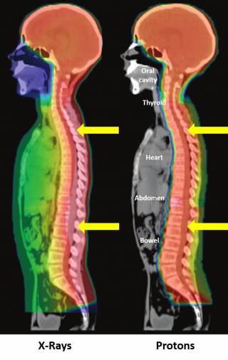
Treatment plans (craniospinal and ewing sarcoma) highlighting an x-ray vs proton comparison
In December 2021, University College London Hospital treated its first patients using Proton Beam Therapy (PBT). One of only two NHS proton beam centres in the UK, the new centre at UCLH brings together some of the world’s leading specialists in complex cancers and will drive forward research into what remains a relatively new treatment.
The advantage of proton beam therapy over conventional radiotherapy is that the bulk of the energy is delivered directly to the tumour with no exit dose. This limits the impact on the surrounding healthy tissue, leading to fewer side-effects and reduced long term consequences, including the risk of a secondary malignancy. Patients treated with PBT range from very young children to adults who have hard to treat cancers. These may be tumours in the brain, on the spine, or near the reproductive organs, where it is particularly important to protect the surrounding tissue. Around a third of the patients will be children and teenagers.
UCL Medical Physics and Biomedical Engineering researchers Professor Gary Royle and Professor Maria Hawkins and their team are working in partnership with UCLH to advance the future of cancer therapy through their research on proton beam therapy and in the development of clinical trials.
Find out more on PBT at UCLH and about the UCL Proton Therapy Research Group.
UCL Medical Physics and Biomedical Engineering, in partnership with UCLH and IPEM will be holding a public lecture on Proton Therapy in Spring 2022, details to follow.
Proton Beam Therapy Research presented at STEM for Britain 2022

Best of luck, Adam! We can’t wait to see your final poster and hear all about the event!