The Department has a long history and we have collected a range of instruments which now form a museum. It's on the third floor of the Malet Place Engineering Building.
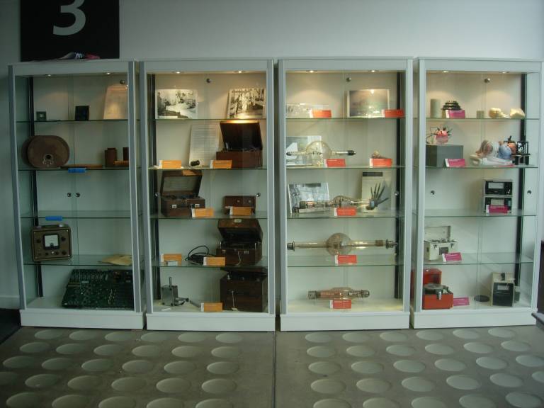
Professor Frank Farmer and his dosemeter
Frank Farmer worked in the Middlesex Hospital Medical Physics Department from 1940 to 1945. During the Second World War, he was part of a consortium of physicists who covered hospitals in London. Much of their work involved handling radium, the first widely used radioisotope. Frank Farmer realised that instruments used for measuring dose were inadequate and began to develop an improved dosemeter.
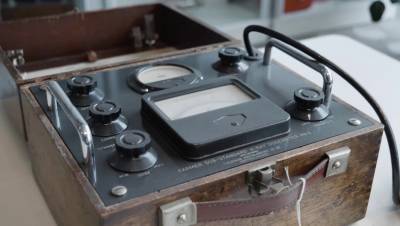
The dosemeter he developed measures electric charge extremely accurately using an arrangement which compensates for drift and systematic errors. First, a voltage is applied across an ionisation chamber. The inner electrode of the ionisation chamber is attached to an electronic valve. The current flowing in the anode of the valve depends on the voltage across the ionisation chamber. Irradiating the ionisation chamber leads to a drop in voltage proportional to the dose received. This then reduces the current flowing in the anode of the valve. An opposing voltage is applied to the valve so that the current returns to its initial value. That voltage is a direct measure of x-ray exposure.
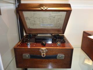
This figure shows the Farmer-Baldwin Dosemeter. In the first prototype, the ionisation chamber is charged using a needle electrode. In the second type, the ionisation chamber is connected with a cable which provides charging and readout. There is also a copy of the first commercial product, and another type which uses mains power instead of a battery.
The Farmer-Baldwin Dosemeter became the most widely-used method of measuring dose accurately. They were commonly used as a secondary standard (or a sub-standard), which means that the instrument has been directly calibrated against the UK’s primary standard. Any other local instruments would be calibrated against the secondary standard, so the calibration can always be traced back to the primary standard.
X-ray generation
The first diagnostic x-ray image in the Middlesex Hospital was taken in 1896, the year following the discovery of x-rays by Prof Wilhelm Roentgen. UCL's first x-ray was taken in the same year by Prof Norman Collie. We have a replica of the first x-ray tube used in the Middlesex Hospital.
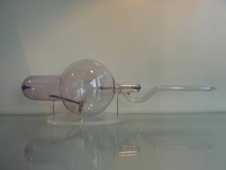
The image above is an early x-ray tube known as a Crookes tube. It was made by Newton & Co. in London, and probably dates from around 1900. Its cathode (inside the tube on the left hand side) has broken off. It is a modification of the kind of tube which Roentgen used to discover x-rays. This type of tube was used from the discovery of x-rays through to the 1920s.
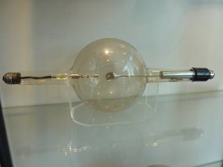
This is a later design of x-ray tube in which electrons emitted from a heated cathode, as in modern devices. Some damage to the anode can be seen, suggesting that this tube was used in the hospital. It probably dates from 1930-1950.
Our display contains a number of x-ray tubes used for diagnosis and radiotherapy, as well as a large valve used for rectifying the high voltage supply required for x-ray generation.
Opening of X-ray Department 1935
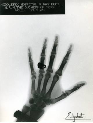
The Duchess of York, later to become the Queen Mother, opened the new Middlesex Hospital x-ray department on 29th May 1935. As a memento of her visit, she had a x-ray image taken of her left hand. The image is reproduced below.
Physiological measurement
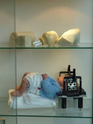
We have a display of some of the tissue-equivalent phantoms and demonstration objects used for optical tomography studies. The image above shows various anatomical test objects and a simulated optical tomography experiment which employs an optical probe holder used to image the newborn baby brain.
We also have a portable electrocardiography (ECG) system which has a paper chart recorder. It was used in UCL Hospitals, and was donated by the family of Clarissa Morse who died aged 4 on 11th January 1961.
Researchers in this department developed the first catheter tip oxygen sensors, which measured blood oxygen concentration in babies. We have some of the original modules used to power and record from these sensors.
Prof John Clifton, who later became Head of Department, designed a Grenz ray chamber which we have in our display. It is used for measuring the dose from very low energy x-rays (10-50 kV).
Thanks
We would like to thank Prof. John Clifton, a previous Head of Department, who provided advice on the content of the museum, and Katie Kilmartin, Sussanah Chan and Jayne Dunn from UCL's Museums and Collections.
 Close
Close

