 Close
Close

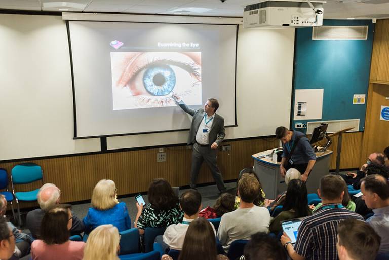
Professor Pearse Keane explains how using artificial intelligence (AI) to investigate the eye's retinal tissue can help diagnose dementia.
What is your primary field of research?
I’m a consultant ophthalmologist at Moorfields Eye Hospital and a Professor of Artificial Medical Intelligence at UCL Institute of Ophthalmology.
Although I’m not a computer scientist or engineer, I lead a multidisciplinary research group which aims to develop and apply artificial intelligence in healthcare, both in the UK and globally, using ophthalmology as an exemplar. We cover the full spectrum of research in clinical AI, going “from idea to algorithm” and “from code to clinic”.
What can detailed images of the inside of the eye tell us about dementia at large?
For more than 100 years, doctors have known that the eye can be used as a window to the rest of the body. This makes sense because the retinal blood vessels are the only microvasculature that can be directly visualised in living human subjects.
And similarly, the retinal tissue is the only part of the central nervous system that can be directly seen without removing part of the skull. It’s an approachable part of the brain! From a number of studies, we have evidence showing that signs of degeneration in the retinal tissue may be seen in patients with dementia.
How have developments in artificial intelligence (AI) and machine learning affected your field of research and how did you become involved in the use of these tools?
Our ability to use the eye as a window to systemic health has been supercharged in recent years by the combination of: 1) big data, 2) the latest advances in artificial intelligence, and 3) advances in retinal imaging such as optical coherence tomography.
This has led to a new term being coined, “oculomics”, to describe an emerging field of study. AI can allow us to find new information hidden within retinal images that is not currently visible to human experts.
How can these new technologies help detect Alzheimer’s and other disorders years before symptoms appear?
We’re still in the early stages of this research but we hope to find changes in the inner layers of the retina that may indicate early signs of neurodegeneration. We hope that some of these changes may be specific to certain conditions and thus could help with screening for these conditions.
What is AlzEye and how can it help prevention?
The AlzEye study is a research study involving UCL and Moorfields Eye Hospital. In it, we have linked ophthalmic imaging data from Moorfields with a national NHS database called Hospital Episode Statistics (HES).
This means that we can identify patients that have had retinal scans done at Moorfields and who have subsequently developed a range of cardiovascular and neurodegenerative diseases. Dr Siegfried Wagner and I lead this study.
What is the hope for the future in this type of research?
We believe that the latest advances in AI will lead to a new era of scientific discovery. We hope that our work will contribute to this, both for ocular disease and for oculomics.
What is the potential of this work for public health?
If we can find strong signs of systemic disease from eye examinations, it could have huge public health potential.
For example, we know that only a small percentage of people in their 40s and 50s attend their GPs for the NHS Health Check. By contrast, however, the majority of these people will attend their local optometrists for regular eye tests. If we can use such tests to pick up the earliest signs of systemic diseases, it would be transformative!
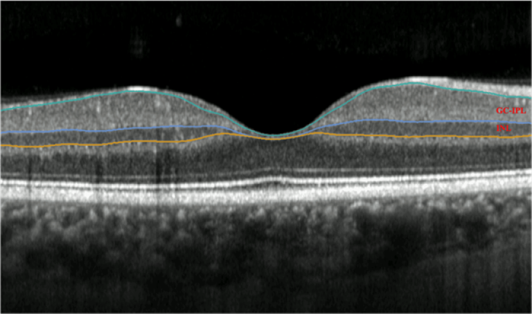
Image: Optical Coherence Tomography (OCT) scan of the retina at the back of the eye, with the layers of the retina highlighted in colour.