 Close
Close

New study reveals data from routine eye scans can be used to detect signs of Parkinson's
21 August 2023
Largest study to date on retinal imaging in Parkinson’s research identifies markers of the condition with the help of artificial intelligence (AI) in eye scans.
A study led by UCL Institute of Ophthalmology's Siegfried Wagner and Pearse Keane, in collaboration with other UCL institutions, has identified markers that indicate the presence of Parkinson’s in patients on average seven years before clinical presentation.
The research findings are presented in a paper published today in Neurology. The analysis of the AlzEye dataset was repeated using the wider UK Biobank database (healthy volunteers), which replicated the discoveries. The use of these two large, powerful datasets has enabled the team to identify these subtle markers, even though Parkinson’s has a relatively low prevalence (0.1-0.2% of the population). Generation of the AlzEye dataset was enabled by INSIGHT, the world's largest database of retinal images and associated clinical data.
The use of data from eye scans has previously revealed signs of other neurodegenerative conditions, including Alzheimer’s, multiple sclerosis and, most recently, schizophrenia, in an emerging and exciting field of research referred to as “oculomics”. Eye scans and eye data have also been able to reveal a propensity to high blood pressure, cardiovascular conditions including strokes, and diabetes.
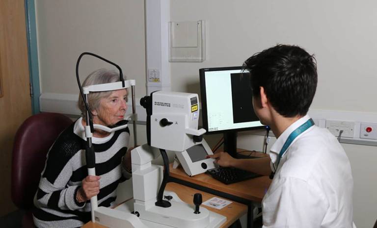
High-resolution images of the retina are now a routine part of eye care, in particular, a type of 3D scan known as ‘optical coherence tomography’ (OCT), which is widely used in eye clinics and high-street opticians. In less than a minute, an OCT scan produces a cross-section of the retina (the back of the eye) in incredible detail, down to a thousandth of a millimetre. These images are extremely useful for monitoring eye health, but their value goes much further, as a scan of the retina is the only non-intrusive way to view layers of cells below the skin’s surface. In recent years, researchers have started to use powerful computers to accurately analyse large numbers of OCTs and other eye images in a fraction of the time it would take a human. Using a type of AI known as ‘machine learning’, computers are now able to uncover hidden information about the whole body from these images alone. Harnessing this new potential is what oculomics is about.
This work is a collaboration between the NIHR (National Institute of Health and Social Care) Biomedical Research Centres at Moorfields Eye Hospital, University Hospital Birmingham, Great Ormond Street Hospital (GOSH), Oxford University Hospital, University College Hospital London and the UCL Great Ormond Street Institute of Child Health. The scope and quality of the research has been maximised through these exceptional NHS research partnerships.
Dr Siegfried Wagner said:
“I continue to be amazed by what we can discover through eye scans. While we are not yet ready to predict whether an individual will develop Parkinson’s, we hope that this method could soon become a pre-screening tool for people at risk of disease. Finding signs of a number of diseases before symptoms emerge means that, in the future, people could have the time to make lifestyle changes to prevent some conditions arising, and clinicians could delay the onset and impact of lifechanging neurodegenerative disorders.
Links
- Research paper in Neurology
- Article on Moorfields Eye Hospital's website
- Article on NIHR Moorfields BRC's website
- Dr Sigfried Wagner's academic profile
- Professor Pearse Keane's academic profile
- INSIGHT hub
- ITV: Study shows eye scans could diagnose Parkinson's disease early by seven years
- BBC: Parkinson's disease could be detected early with AI scans, scientists say
- Guardian: 3D eye scans at opticians could identify those at risk of Parkinson’s, study finds
- Evening Standard: Eye scans detect signs of Parkinson’s disease up to seven years before diagnosis
- Evening Standard: AI-powered eye scan could help detect Parkinson’s disease – study