 Close
Close

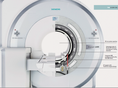
The high resolution and sensitivity of PET/MRI captures minute details and produces superior three-dimensional images available on the 3T system. The simultaneous image capture of the PET and MRI scans eliminates the potential imaging problems caused by involuntary patient movement, such as breathing and muscle relaxation. When used for anatomical imaging, the radiation dose for patients is half that of the next best technology.
PET/MRI
The combined scanner reduces the need for patients to come for multiple visits. Patient diagnosis is faster because imaging and overall information available from the scan is better. The magnet is at 3T field strength based on Siemens Verio model.
Current clinical protocols include but are not limited to:
Whole body 18F-FDG with 3T MRI structural and functional imaging
Neuroimaging 18F-FDG with 3T MRI structural and functional imaging for epilepsy and dementia.
Current research projects underway include:
Differential diagnosis of dementia 18F-FDG with 3T MRI structural and functional imaging.
The role of PET/MRI in radiotherapy planning for meningioma using 68-Ga DOTATATE and 3T MRI structural and functional imaging. Biomarkers for angiogenesis and hypoxia using 18F-FDG and 3T MRI advanced functional imaging.
Please contact Prof. Ashley Groves for prospective research projects or Dr. Jimmy Bomanjii for clinical referrals.
Please contact Dr. Anna Barnes for technical details or MRI sequence development.