 Close
Close

First EBS image: The data that could revolutionise histology
24 March 2021
The images promise a new era in histology, where entire organs – even entire bodies – can be biopsied without lifting a scalpel.
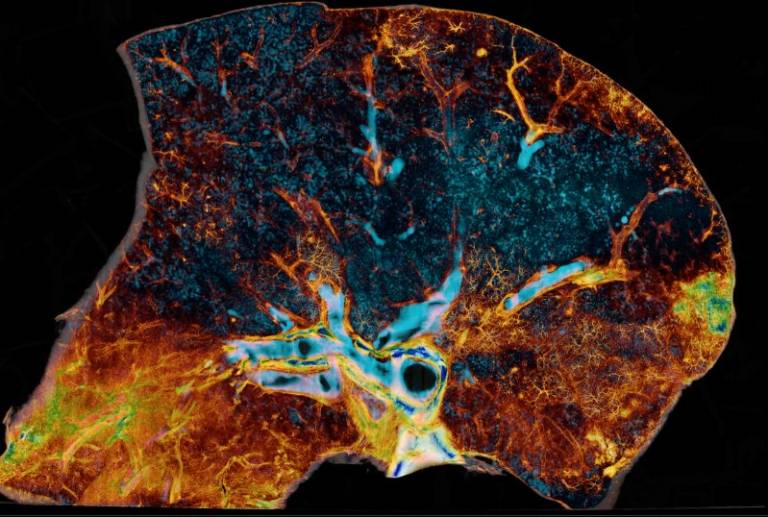
At present count, Peter Lee and his medical teams in Germany and France have shown the images of a COVID-19-injured lung to more than 100 physicians and biologists worldwide. Every time, he witnesses the same reaction. “First they say, ‘Wow!’ And then they scratch their heads. They’ve never had to think about how to use this kind of information – it’s not existed before.” The images – including the one shown here – are the first data to be publicised after the ESRF’s EBS upgrade, and it is easy to see why people are impressed.
Even to the untrained eye, the level of detail contained in them is astonishing. At the click of a mouse, everything is visible, from the entire organ and its major airways, all the way down to the alveoli and the finest micro-vasculature. But to medical scientists, the images are way more than just aesthetically pleasing. They promise a new era in histology, where entire organs –even entire bodies –can be biopsied without lifting a scalpel, unmasking the complex and interconnected pathologies of disease.
The preliminary data are so striking that in December, Professors Peter Lee and Rebecca Shipley, together with Drs Tafforeau (ESRF) and Walsh, together with their German Medical collaborators, were awarded a $1m (£720k) grant from the Chan Zuckerberg Initiative, a philanthropy founded by Priscilla Chan, a paediatrician, and her husband Mark Zuckerberg, the founder of Facebook. By 2023, if all goes to plan, Lee and his colleagues will imaged an entire human torso in three dimensions, non-destructively, at hundreds of times better resolution than has previously been achieved.