 Close
Close

Imaging improved by scrambling X-rays
21 July 2014
X-ray phase-contrast imaging can provide
high-quality images of objects with lower radiation doses. But until now these
images have been hard to obtain and required special X-ray sources whose
properties are typically only found at large particle accelerator facilities.
Using a laboratory source with unprecedented brightness, scientists from the Technische Universität München (TUM) and the Royal Institute of Technology in Stockholm (KTH), as well as Dr Pierre Thibault (UCL Physics & Astronomy), have demonstrated a new approach to get reliable phase contrast with an extremely simple setup.
X-ray phase-contrast imaging is a method
that uses the refraction (bending) of X-rays as they pass through a specimen instead of their
attenuation (dimming), which often produces images of much higher quality.
In their new study, the scientists have now developed an extremely simple setup to produce X-ray phase-contrast images. The solution to many of their difficulties may seem counter-intuitive: scramble the X-rays to give them a random structure. These so-called 'speckles' encode a wealth of information about the sample as they travel through it. The scrambled X-rays are collected with a high-resolution X-ray camera, and the information is then extracted afterwards.
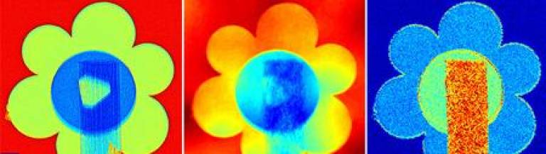
Using their new technique, the researchers have demonstrated the efficiency and versatility of their approach. "From a single measurement, we obtain an attenuation image, the phase image, but also a dark-field image," explains Dr Irene Zanette (TUM), lead author of the publication. "The phase image can be used to measure accurately the specimen's projected thickness. The dark-field image can be just as important because it maps structures in the specimen too small to be resolved, such as cracks or fibers in materials," she adds.
The source's high brightness is also key to these results. "In the source we used a liquid metal jet as the X-ray-producing target instead of the solid targets normally used in laboratory X-ray sources," says Tunhe Zhou from KTH Stockholm, project partner of the TUM. "This makes it possible to gain the high intensity needed for phase-contrast imaging without damaging the X-ray-producing target."
To obtain all images at once, an algorithm
scans the speckles and analyzes the minute changes in their shape and position
caused by the specimen. "Our refined speckle-tracking approach is quite
robust" says Dr Pierre Thibault, from UCL. "We have also introduced
a phase integration step that converts
the deflection angles into accurate quantitative phase images."
Notes
- The research is published in the journal Physical Review Letters in an article entitled 'Speckle-based X-ray Phase-contrast and dark-field imaging with a laboratory source'
Related links
High-resolution images
Attenuation, phase-contrast and dark-field images
Intensity landscape of X-ray speckles
These images can be reproduced freely for the purposes of news reporting and discussion. For other queries, please contact Dr Irene Zanette (Technical University of Munich, Germany), on irene.zanette@tum.de or by phone on +49 89 289 10802.
Science contact
Dr Pierre Thibault
UCL Physics & Astronomy
020 7679 1558
p.thibault@ucl.ac.uk
Media contact
Oli Usher
UCL Faculty of Mathematical & Physical Sciences
020 7679 7964
o.usher@ucl.ac.uk
Open Days

The Faculty participates in a number of open days throughout the academic year, including the UCL Undergraduate Open Days and the UCL Graduate Open Day.

Friends of Out@UCL
Professor Ivan Parkin -
Dean, UCL Faculty of Mathematical and Physical Sciences
"I fully support the aims of the Friends of Out@UCL campaign. I have
personal experience of the need for such a campaign and the
difficulties that the LGBTQ+ community face." Read more...
Snapshots from Space History

Online exhibition of historic space photos from the faculty's planetary science archives.