Close
Close

IHE Summer Studentships 2023
15 February 2023
UCL Engineering and the Institute of Healthcare Engineering are offering multiple summer studentships at the Royal National Orthopaedic Hospital, UCLH/UCLH BRC, and Moorfields BRC for UCL undergraduates.

Please note the application for student applications has been extended to Friday 14 April.
The IHE Summer Studentship Scheme will give undergraduate students an exciting opportunity to shadow clinicians and take part in a related research project at the Royal National Orthopaedic Hospital (RNOH), UCLH/UCLH Biomedical Research Centre and Moorfields Biomedial Research Centre. The projects will be jointly supervised by a clinician and a UCL academic.
Overview
Eligibility
The studentship scheme is open to UCL Engineering undergraduate students in their second year and above.
Dates and duration
Studentships will run for 8 weeks from June through September with the exact dates to be agreed between supervisors and students for each project. It is expected that the internships will be taken partly or fully in person.
Finances
Funding for 8 weeks for the student (that is 36.5 hours/week *£11.95/hour * 8 weeks, i.e. £3,585) + £200 stipend for the project.
What will a typical project look like?
In the first 3 weeks, students will have exposure to the clinical setting and interaction with clinical teams. The remaining 5 weeks will be focused on their selected project. Ideally, the core of the project should be completed by end of week 7, using week 8 for a write-up, handover, and dissemination.
Information for clinicians and UCL staff
Project proposals are now closed.
Information for UCL students
How to apply
Applications for the scheme are now open. Deadline: 11.59 PM, Friday 14 April 2023.
To apply for the scheme, you must submit:
- Your CV (2-pages maximum). Check out this advice about formatting your CV from the Complete University Guide.
- A cover letter (1-page maximum), and indicate your 1st and 2nd choice of projects with a justification of why you are suitable for each (200 words per project).
- A statement of endorsement from your personal tutor or another UCL academic staff member who knows you personally with their contact details. This person may be contacted if there are any issues (e.g. wellbeing or engagement) during your studentship, so please make sure they are aware of this.
- Submit your details via this form.
We are looking for applicants with a history of academic excellence, motivation, and leadership, as well as a convincing case of suitability for their chosen project.
Please see the project details below, or download the full list.
Important note:
Some additional projects might be included until the deadline for students’ submission. You can access always the most updated project list via this website. The current project list is a good representation of the projects that are on offer and so you can submit from now. But if you want to wait until closer to the deadline, at least you should prepare a draft of your application considering these current projects to ensure you are ready to submit at any time.
- Project 1: Design of force measuring apparatus for investigating screw compression, RNOH
Clinical unit: Royal National Orthopaedic Hospital (RNOH) - Foot & Ankle Unit
Lead clinicians: Mr Karan Malhotra
Project outline: In orthopaedics the screw is a critical device. It is a device which converts rotation force into a linear compression and must maintain that compression and resist pullout and distraction. To achieve these goals various screw designs have been developed varying the pitch, thread depth and thread flank configuration. However it is not known which screw types generate optimal compression. In order to measure and compare screws a purpose designed apparatus will be required to measure compression force generated and pull-out strength. This project will be aimed at designing and building an apparatus for measuring force generation by screws (compression). This may later be used as part of a larger project aimed at investigating and comparing various screw designs and is a critical first step.
Outputs: Designing an apparatus for measuring force generation by screws (compression) Skills and clinical observation opportunities students will gain from this project: Students will have the opportunity to join consultant surgeons in clinic and theatre to see the role of screws / metalwork in clinical practice, and to learn about the biomechanical and biological aspects. There will be ample opportunity to discuss clinical applications and discuss design.
Pre-existing skills needed for this project: NA
- Project 2: Artificial Intelligence for Optimising the Design and Placement of Patient Specific Posterior Pedicle Screws in Scoliosis Surgery, RNOH
Clinical unit: Royal National Orthopaedic Hospital (RNOH)
Lead clinicians: Prof Deepak Kalaskar - Professor of Bioengineering (Department of Ortho and MSK Science, Division of Surgery & Interventional Sciences) Mr Sangan Patil - Spinal Surgeon
Project outline: Scoliosis surgery involves the insertion of screws (pedicle screws) in the spine. Current techniques to insert these screws are not completely accurate. Even in international experts’ hands, 25-30% of the screws are misplaced. Misplacement of screws has a high risk of bone weakening, injuries to the spinal cord, nerve roots or blood vessels. Getting screw placement wrong has long term health implications for young patients, including lifelong disability. More accurate methods use computer navigation or image guidance techniques. However, these involve using more radiation (X-rays, CT scans) before and/or during the surgery. Surgeons and parents are concerned regarding the long-term effects of ionising radiation in young patients. This project will address this unmet health need by developing efficient pre-surgical planning workflows for the accurate placement of pedicle screws based on artificial intelligence and machine learning. This will remove the need for additional imaging with ionising radiation and optimise the surgical planning process.
Outputs: Primary aim of this study is to develop machine learning architectures to support accurate placement of patient specific posterior pedicle screw insertion guides for scoliosis surgery from CT imaging. The project outcomes will minimise the need for additional imaging and after integrated in an established workflow, will accelerate the entire pre-operative planning process including the design and manufacturing of pedicle screw guides.
Pre-existing skills needed for this project: Specific skills are not required but an overall understanding of CT/MRI imaging and spinal structure would be beneficial
- Project 3: Investigating myopia in children and evaluating novel treatments: a long-term approach to short-sightedness, Moorfields
Clinical unit: Children's Service at Moorfields Eye Hospital NHS Foundation Trust
Lead clinicians: Dr. Annegret Dahlmann-Noor
Project outline: Short-sightedness, more formally called myopia, is increasing in children in the UK, and around the world. Aside from the cost of glasses and the day-to-day nuisance of wearing glasses, there is also a more severe side to myopia in children. Early onset myopia is associated with extreme vision loss in later life. Myopia, predicted to affect half the world population by 2050, is set to become a leading cause for irreversible sight loss. Reducing the costs of childhood myopia now and saving their vision in the future is an extremely important problem for clinical care and new technology development. The purpose of this project is to investigate changes to the inner structures of the eye that may be associated with the severity of myopia. The student will be given an opportunity to shadow the clinician at Moorfields Eye Hospital and spend time in the clinic. The focus of the student’s contributions will be on developing image processing tools that can be used to analyse clinical ocular images acquired using a technology called optical coherence tomography (OCT).
Outputs: This project will be led collaboratively by the clinician at Moorfields Eye Hospital and the academic partner at UCL. The student outputs will provide technical contributions towards answering the clinical questions and providing mechanistic insights to myopia.
Pre-existing skills needed for this project: Yes – introductory scientific programming with MATLAB (preferred) or Python.
- Project 4: The Analysis of Magnetically Controlled Growing Rods for the Treatment of Children with Early Onset Scoliosis, RNOH
Clinical unit: The Royal National Orthopaedic Hospital (RNOH) Implant Science Centre
Lead clinicians: Mr Johann Henckel
Project outline: The Implant Science Centre, based at the Royal National Orthopaedic Hospital (RNOH) is the largest centre in Europe for the analysis of implants retrieved from patients. The centre has collected over 10,000 spine, hip and knee implants from patients across 29 countries and published over 150 papers on this subject.
Outputs: The student will generate data relating to the analysis of the MCGRs in this study. The analysis of this, in the context of available literature and associated data relating to these patients, will be written up as a final report. It is expected that the data will be used to develop a manuscript for submission to a peer-review journal.
Pre-existing skills needed for this project: Student should have an interest and background in engineering, bioengineering, biomechanics in orthopaedics and multidisciplinary research, or a similar discipline.
- Project 5: Stereotactic Body Radiotherapy for Liver Cancer : An Evaluation of Normal Tissue Dosimetry, UCLH
Clinical unit: UCLH Radiotherapy Department
Lead clinicians: Prof Maria Hawkins Dr Douglas Brand
Project outline: Hepatocellular carcinoma (cancer of the liver) affects around 6000 patients in the UK each year, with higher incidences seen in countries with endemic viral hepatitis. For patients who have disease localised to their liver, stereotactic body radiotherapy (SBRT) offers a potentially curative treatment modality for those where surgery is not feasible. This involves delivery of large doses of highly targeted radiation, delivered typically over 5 days.
The critical issue in radiotherapy treatments for cancer is the therapeutic ratio: describing the balance between the probability of tumour control and the probability of experiencing normal tissue toxicity. In routine practice this is achieved through use of a target tumour dose and dose constraints applied to normal tissues. For patients with liver cancer a key concern is toxicity occurring in their normal liver, which may be damaged due to alcohol use or other causes of cirrhosis (which predispose to liver cancer). Newer radiotherapy delivery methods such as proton beam therapy offer a potential method to reduce the dose to the liver, but the precise benefit expected is uncertain.
In this project the candidate will have access to the UCLH radiotherapy system to examine clinical/tumour factors and doses to normal tissues amongst patients receiving SBRT for liver cancer. Depending on interests, there will be the possibility to develop models to predict dose, along with multivariate outcome prediction modelling. Work from this will help to contribute to the development of a new national trial aiming to test proton radiotherapy for liver cancer.
Outputs:
1) Completion of a clinical service evaluation
2) Present findings as a conference abstract (international level preferred)
3) Write up findings into a full-length research paper
4) Potential to develop work into longer format research project in the future (e.g. masters or doctoral level project)
Pre-existing skills needed for this project: Some understanding of medical imaging types and data analysis would be helpful but not essential.
- Project 6: Studying the spino-pelvic relationship prior to total hip replacement: Co-registration of two image modalities, RNOH
Clinical unit: Surgical Technology Laboratory, Royal National Orthopaedic Hospital, Stanmore
Lead clinicians: Mr Johann Henckel
Project outline:
Significant variability has been reported for pelvic orientation in patients with hip arthritis with almost all individuals having a unique pelvic tilt which changes over time and with daily activities. Understanding the magnitude of these changes is important as it influences surgeon’s decision regarding the optimal implant orientation for the patient (e.g. to reduce the risk of dislocation), however this has been challenging to accurately measure using standard imaging modalities.
Some of the main complications associated with hip replacements include dislocation, impingement, reduced range of movement and accelerated implant wear. A better understanding the impact of pelvic orientation on implant function will improve surgical planning and execution as well as outcome. Although most hip dislocations occur in either standing or sitting position, the safe zone for implant position is currently defined using the supine position.
Computed Tomography (CT) is becoming the gold standard imaging for 3D surgical planning and is almost always performed with the patient in the supine position (non-functional). Biplanar radiography, EOS, offers upright full-length imaging to accurately measure patient’s anatomy. However, it does not offer the 3D information that CT offers.
The aim of the project is to study the magnitude of variation in pelvic tilt from lying to standing in patients undergoing hip replacement by seeking to accurately co-register these two imaging modalities (CT and EOS) using standardised coordinate systems. The change in pelvic orientation form lying (non-functional posture) and standing (functional posture) will inform planning of implant component orientation (e.g. acetabular cup position) to optimise function, range of movement and implant longevity.
Outputs: The change in pelvic orientation form lying (non-functional posture) and standing (functional posture) will inform planning of implant component orientation (e.g. acetabular cup position) to optimise function, range of movement and implant longevity. We would expect the results of this study to be written up as a manuscript for peer-review.
Pre-existing skills needed for this project: Student with interest and background in bioengineering, biomechanics in orthopaedics, multidisciplinary research. The project will include 3D image analysis and the use of state-of-the-art software solutions for image segmentation and registration.
- Project 7: Using Cardio-Pulmonary Exercise Test (CPET) to predict post-surgery outcomes, UCLH
Clinical unit: UCLH - Human Pharmacology and Physiology Laboratory
Lead clinicians: Dr John Whittle, Consultant in Anaesthesia, Critical Care + Perioperative Medicine, UCLH
Project outline: From a joint collaboration between the UCLH perioperative unit and the WEISS Centre, we propose a project that will aim to create the foundation for a machine learning approach to determine the odds of developing complications after surgery and finally predict the long term mortality.
A surgical operation represents an enormous physical and mental challenge for humans, like a strenuous run or hiking, with a complex impact on the quality and quantity of life during and after the surgery. Every year more than 350 Million surgery operations are conducted around the world, and future predictions show a potential increase up to 500 million/years by 2050. Concurrently, the human population is globally getting older and the amount of people with relevant medical conditions (Arterial hypertension, Diabetes, Obesity, etc.) is increasing. Thus a population that is getting every day more complex leads to a higher risk of developing complications and adverse outcomes from surgery.
Nowadays, the main clinical approach is the clinical use of the Enhanced recovery after surgery (ERAS) protocols. The ERAS are multimodal perioperative care pathways designed to achieve early recovery after surgical procedures by maintaining preoperative organ function and reducing the profound stress response following surgery. But despite this innovative approach, 1 in 5 patients develops serious perioperative complications. Currently, anaesthetists and clinicians are not able to predict in advance the development of these complications and clinical scores lack the ability to stratify the patients before the surgery. But, again, technology is on our side.
We can currently use the cardio-pulmonary exercise test (CPET) to obtain an enormous amount of extremely interesting data from patients at rest and under stress, mimicking the surgical stress and recording their physiological responses. This response to stress is strictly correlated with the development of future complications after surgery.
Furthermore, these data minutely describe how our cardiovascular system and our metabolism adapt to physical stress, but its analysis is complex and is it not possible to be done in real-time by humans, but could be done using a multimodal machine learning approach. With a machine-based approach we aim to be able to understand to describe the different phenotypes of human physiology and their correlation with surgical stress and perioperative outcome.
Finally, the aim of this project is to develop the first fundamental steps of a future clinical tool to help clinicians to better understand the risk for patients undergoing surgery, creating the base for future medical interventions.
The project will be developed over two months, with the first 4 weeks phase of clinical introduction and development of the database and a second 4 weeks phase of creating the model with the support and assistance of clinicians and engineers lead.
- Project 8: Optimastion of stimulation parameters for neuromodulation in people living with Spinal Cord Injury, RNOH
Clinical unit: London Spinal Cord Injury Centre, RNOH.
Lead clinicians: Lt Col David Baxter
Project outline: Spinal Cord Injury (SCI) is a life-changing event, permanently affecting the control of limbs and organs, causing loss of sensation, unbalanced autonomic function, neuropathic pain, and many more debilitating consequences. Neuromodulation, in the form of electrical stimulation applied over the spinal cord, has been trialled as a rehabilitative intervention in people with SCI with remarkable reports of recovered function of lower limb muscles. Benefits to autonomic functions including cardiovascular function and bladder, bowel and sexual function have also been reported. One of the most challenging aspects in the clinical application of neuromodulation after SCI, is parameter optimisation. Epidural electrode arrays typically contain up to 16 electrodes and stimulation can be applied at a wide range of amplitudes, pulse widths and frequencies resulting in trillions of possible settings.
In this project, the student will investigate:
1. Optimal stimulation amplitudes and frequencies for facilitating or inhibiting corticospinal excitability. Healthy volunteers will be recruited and receive short interventions of non-invasive spinal cord stimulation (SCS) with various stimulation amplitudes and frequencies. Electromyography (EMG), transcranial magnetic stimulation (TMS) and electrical nerve stimulation will be applied before and after the intervention to assess the effects on spinal and corticospinal excitability.
2. Lower limb mapping procedures for spatial optimisation. Lower limb mapping will be carried out in individuals with SCI who already have implanted epidural stimulators.
The aim is to identify a configuration that activates all lower limb muscles, at a similar stimulation amplitude. Lower limb EMG will be used to measure responses to low frequency stimulation applied using different electrode configurations with a range of stimulation intestines (recruitment curves). The student will analyse the data collected, conduct a literature review of mapping procedures used by other groups, and initiate a standard mapping protocol that can be used in future for new patients with SCI receiving epidural stimulators.
Outputs: The student will analyse the data collected, which we expect to lead to a conference presentation and/or journal publication. The student will also initiate a protocol for lower limb mapping in people with SCI who have epidural stimulators implanted. We expect this protocol to form part of the guidelines we are developing for the application of neuromodulation after SCI, which will be shared with other SCI centres throughout the UK.
Pre-existing skills needed for this project: A student with knowledge of anatomy and physiology or an intercalating medic would be most suited to the project.
- Project 9: Clinical Training Phantoms for Endobronchial Ultrasound and Bronchoscopy
Clinical unit: Thoracic Medicine
Lead clinicians: Dr Ricky Thakrar Dr Neal Navani
Project outline: Lung cancer is a leading cause of cancer deaths worldwide. Diagnosis and staging are often performed using endobronchial ultrasound and transbronchial needle aspiration (EBUS-TBNA) [1], a procedure that involves using an ultrasound-equipped flexible bronchoscope to navigate the lungs and take biopsies. Mastery of EBUS-TBNA requires extensive training, but in the UK, there is no formal training program, and traditional apprenticeship methods, where a novice learns on patients, carry the risk of harm [2]. International guidelines recommend simulation training and validated skills assessments to objectively evaluate proficiency [3]. High-fidelity EBUS simulators have been proven to be effective in training [4]. However, commercially available simulators have fundamental limitations: they lack key anatomical features, unrealistic ultrasound properties, and incompatibility with needle biopsies. These limitations and high costs make them less suitable for widespread use in EBUS training. The unmet health need addressed here is the lack of high-fidelity, cost-effective models for training clinicians in EBUS. This has direct implications for patient health, as clinical training is key to optimising procedure outcomes.
University College London have developed a novel framework for creating lifelike clinical training phantoms for EBUS procedures. Using advances in inexpensive soft tissue-mimicking materials and 3D printing technologies [5], the engineering team have fabricated anatomically accurate models with a non-toxic material that presents highly realistic ultrasound images, thereby allowing for effective training in lymph node aspiration. Our initial tests indicate that the first-generation phantoms are highly promising for training with accurate airway structure and lymph node anatomy. They still need to have vascular structures configured into the phantom to make them more realistic, but this will add an extra layer of complexity in the fabrication process and require translation of live bronchoscopy images and imaging data to create a lifelike model. This studentship has been designed to be achievable within a timeframe of 8 weeks, offering the successful candidate ample opportunities for clinical engagement. The program involves spending the first weeks working closely with both the clinical and engineering team, observing bronchoscopy procedures, with the aim of developing a new generation of phantoms that are anatomically accurate and can generate highly realistic ultrasound images of vessels and lymph nodes. They will also design and conduct a validation study of the phantom, through evaluation by EBUS experts, who have extensive experience in training apprentices on real patients. This validation study is crucial as it will determine whether the phantom can adequately replace and supplement traditional training methods. The final week will provide time to write up the data with the goal to have a fully developed and validated phantom that can integrate into clinical practice, improving the quality of training in bronchoscopy procedures.
Outputs:
1) to fabricate a new generation of endobronchial ultrasound phantoms in collaboration with the Wellcome-EPSRC Centre for Interventional and Surgical Sciences (WEISS);
2) to determine the feasibility of using a phantom as a clinical training simulator for EBUS-guided transbronchial needle aspiration (EBUS-TBNA) through an evaluation study conducted by expert clinicians. The primary outcome measured will be unbiased scoring of the models performance, with the goal of achieving a reliable inter-rater agreement.
- Project 10: Design of a biomedical device to enhance safety and efficacy of ophthalmic topical medications
Clinical unit: UCL Institute of Ophthalmology & Moorfields Eye Hospital NHS Foundation Trust
Lead clinicians: Paul Foster
Project outline: Many ophthalmic medications are produced in a topical format, which are administered to the surface of the eye in a liquid drop. These medications have their actions through direct absorption into the ocular tissues. However, the drop will drain from the surface of the eye down the lacrimal canaliculi, into the lacrimal sac, and thence into the nose down the nasolacrimal duct. Once in the nasal cavity, the medication may become aerosolised and inhaled into the lungs where it has rapidly absorbed into the systemic circulation. Some of ophthalmic medications (those used for management of intraocular pressure and control of glaucoma) may have significant systemic side-effects. Notable among these are the topical beta blockers such as timolol and the adrenergic agonist such as brimondine and apraclonidine. These can have a potent effect on respiratory and cardiovascular function.
To enhance the efficacy of the medication, and to prevent systemic absorption, causing potentially serious unwanted side-effects, patients are advised to perform a procedure called punctual occlusion. This procedure is accomplished by pinching either side of the nose for up to 5 minutes. It is further enhanced by closure of the eyelids. A combination of these two manoeuvres (a) controls the pumping mechanism which draws fluid off the surface of the eye and (b) blocks drainage of fluid down the lachrymal canaliculi into the lacrimal sac.
This process is rarely followed to its completion and patients find the process of punctual, occlusion onerous. The object of this project is to design a device which can be clipped onto the bridge of the nose, compressing the lacrimal sac and removing the need for digital occlusion of the lachrymal canaliculi. The instigator of this project is a patient of mine (Mr John Fearon).
The project would cover design and prototyping, over one year.
Outputs:
1) to fabricate a new generation of endobronchial ultrasound phantoms in collaboration with the Wellcome-EPSRC Centre for Interventional and Surgical Sciences (WEISS);
2) to determine the feasibility of using a phantom as a clinical training simulator for EBUS-guided transbronchial needle aspiration (EBUS-TBNA) through an evaluation study conducted by expert clinicians. The primary outcome measured will be unbiased scoring of the models performance, with the goal of achieving a reliable inter-rater agreement
How have previous students found the experience?
"My time at the Royal National Orthopaedic Hospital on the IHE Studentship has been an incredible experience and I have learnt a lot. I had the opportunity to work with an amazing team of clinicians and make connections with professionals across the country. I thoroughly enjoyed my time as a summer student, and I am now continuing on as a research assistant on the project part-time!
As someone with a very strong interest in working in a clinical setting, I was really excited to be able to observe in clinics and theatre almost every week, where I was able to speak to spinal sarcoma patients and understand more about the impacts the condition can have on them."
- Vishlesha Vinjamuri (UCL Medical Physics and Biomedical Engineering)
On the other hand, the face-to-face time is improving my practical skills using tools and machines. I am finding the studentship very fulfilling and perhaps one of the most important discoveries that I have made is that I am very clear that after I graduate, I would like to continue to do research in the biomechanics field."
- Alba Morillo Paterson (UCL Medical Physics and Biomedical Engineering)
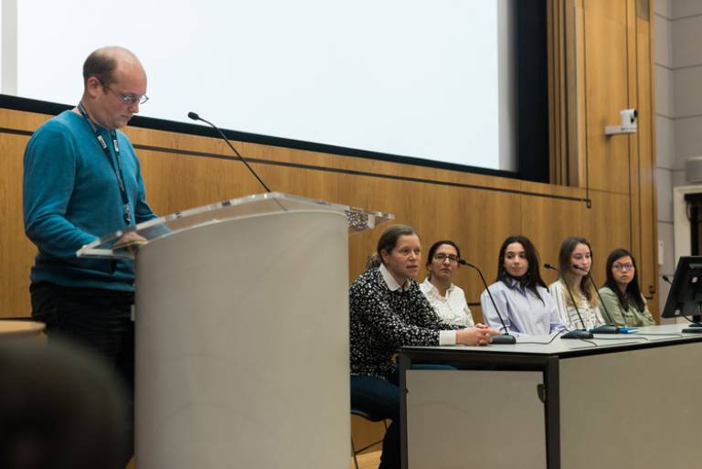
Students from the scheme presented their research project at the IHE's annual research symposium in October 2022.