Location:
Telephone Extension:
Laboratory Manager: Dr Dominic Papineau

This laboratory houses petrographic microscopes, a micro-Raman imaging system, and sample preparation apparatus.
The main petrographic microscope is an Olympus BX51 optical microscope with imaging modes in transmitted light (plane light), cross-polars, and reflected and dark-field. All objectives available are for polished thin sections (5X, 10X, 20X, 50X, and 100X). Targets of microscopic minerals of interest are identified by this technique prior to analysis by micro-Raman.
The micro-Raman in a WITec 300alpha Raman system equipped with a 532 nanometre wavelength laser that can be tuned to between 0 and 45 mW (class 3b). Two gratings of 600 and 1800 lines per milimetre are available for analysis and give spectral resolutions (precision) between 0.1 and 4 cm-1 wavenumbers and bandwidths between 800 and 4000 cm-1. Spectral accuracy is calibrated against an in-house diamond standard and is better than 1 cm-1. Three possible optic fibers with diameters of 25, 50, and 100 micrometres that can be used to sample the scattered photons (these set the pinhole size for confocality). The large area scan function of the WITec software allows users to perform micro-Raman imaging on areas up to 1.5 x 1.5 millimetres in size. A ‘True surface’ add-on further enables the analysis of smooth three-dimensional surfaces.

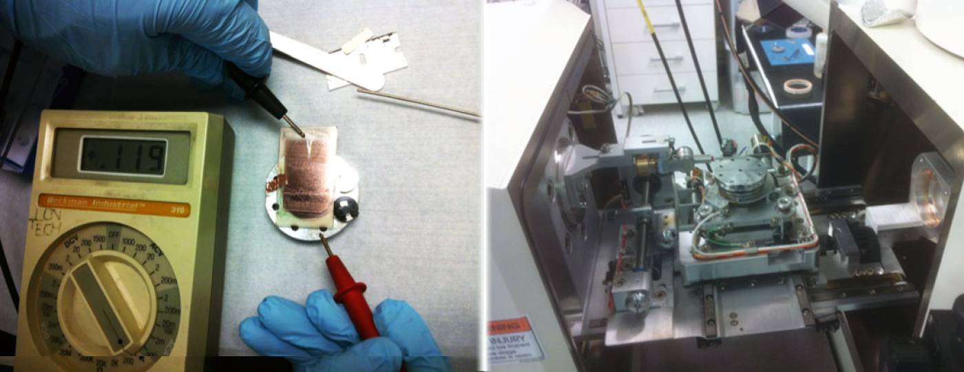
Image:(Left): To ensure good electronic conductivity, we measure the resistance of gold-coated polished thin section prior to insertion into the sample chamber for nano-fabrication. ; (Right) Opened sample chamber of the Leo 1540 XB FIB with the rounded stage in the centre and the micro-manipulator arm on the left.
We also have access to the nano-fabrication cleanroom of the London Centre for Nanotechnology to perform sample preparation by Focused Ion Beam (FIB – using Ga, Ne, and He beams). In the chamber of the Carl Zeiss 1540 XB FIB, a Kleindiek micro-manipulator was recently installed to perform in situ lift-out of nano-fabricated lamellae of specific mineral assemblages. Using the Zeiss Orion NanoFAB, we can thin down our lamellae with atomically-flat surfaces, which is an ideal sample preparation for various micro-analytical techniques.
 Close
Close

