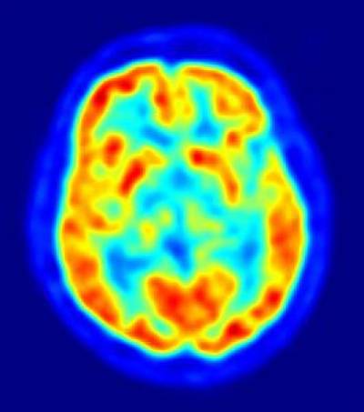
Positron emission tomography (PET) imaging involves injecting a radioactive tracer into a patient and then imaging where this radioactive tracer accumulates. There are numerous tracers that can be used with PET, dependent on which type of function one wishes to monitor. For example, tracer FDG (fludeoxyglucose) is a radioactively labelled glucose that indicates which parts of the brain are more metabolically active, an indicator of brain function. Newly developed tracers attach to the amyloid plaques that are known to be hallmarks of Alzheimer's disease (AD). Evidence of these plaques in people who do not yet show symptoms of AD is a strong risk factor of developing the disease. These plaques have been observed using PET in healthy people 10-15 years before the disease process begins. In the picture, FDG PET image shows glucose uptake in the brain, red regions show parts of the brain that are more metabolically active, blue regions less so.
 Close
Close


