Elizabeth Gordon, Dementia Research Centre
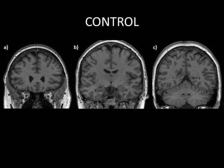
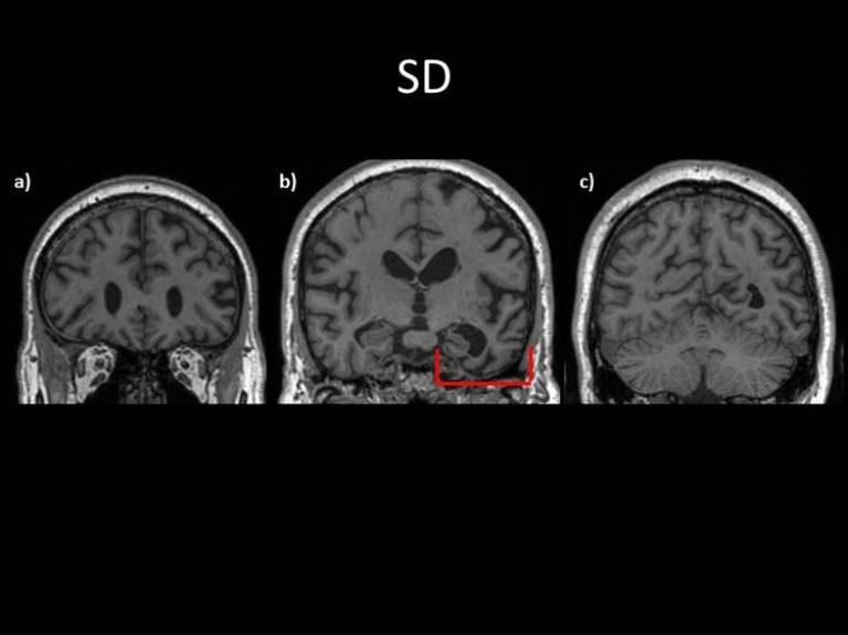
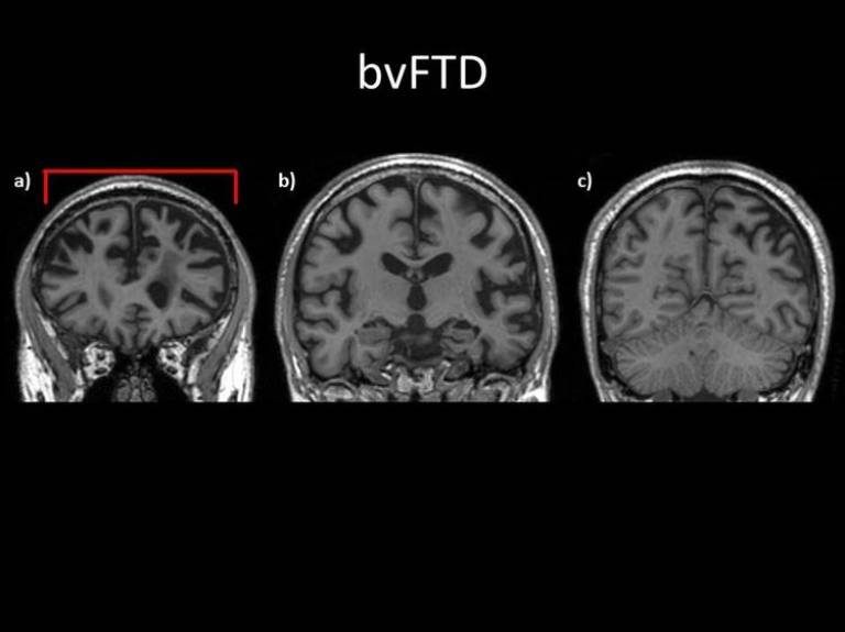
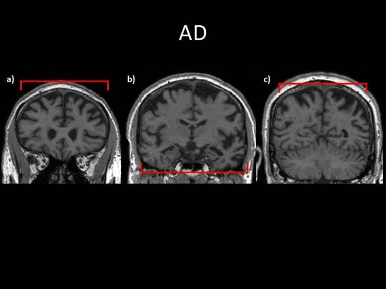
These images are MRI (magnetic resonance imaging) scans from four different individuals. They show a few sections throughout the brain starting from a) the anterior (frontal) regions, moving to b) about half way through the brain and finally c) more posteriorly (towards the back) of the brain.
The first scan shows how the normal healthy brain of a 60 year old looks.

The second image series is taken from someone with semantic dementia. Note the more marked atrophy (shrinking, due to loss of brain cells) of the left hand side of the brain. This is particularly affecting the left temporal lobe (the bottom right of the picture). This part of the brain is heavily involved in knowledge about things and is key for language processing in keeping with the symptoms seen with this disease.

The third set of images are taken from someone with a diagnosis of behavioural variant frontotemporal dementia (bvFTD). While there is more variability within this patient population in terms of atrophy (cell loss) pattern across the brain, in general, there is more pronounced atrophy in the anterior (front) part of the brain with the posterior (back) areas relatively more preserved. These frontal brain regions are involved in co-ordinating and monitoring complex behaviours, personality and planning, which are often primarily affected in bvFTD.

The final images show someone with Alzheimer’s disease. There has been loss of cells all over the brain rather than the focal loss of cells seen in FTD.

 Close
Close

