 Close
Close

How cancer cells communicate shown for first time
18 February 2020
New technology developed at UCL is, for the first time, enabling cancer scientists to analyse the individual behaviour of millions of different cells living inside lab-grown tumours – a breakthrough which could lead to new personalised cancer treatments.
The research, published in Nature Methods, provides new insight into how mutated cancer cells “mimic the growth signals” normally expressed by healthy cells – which allows cancer cells to grow unchecked.
Corresponding author, Dr Chris Tape (UCL Cancer Institute), said: “Our new technology allows us to simultaneously measure the behaviour of cancer cells, healthy cells, and immune cells from mini-tumours.
"This new technique revealed that mutations in cancer cells mimic the growth signals normally provided by cells in the healthy tissue microenvironment. In healthy tissues, signals from the environment are tightly controlled so the tissue doesn’t grow too fast. Unfortunately in cancer, mutations that mimic microenvironment signals are constantly switched on - allowing the cancer to grow unchecked.
"The new technology developed at UCL enabled scientists to observe this phenomenon in minute detail.”
Globally, researchers can now study cancer using mini-tumours, known as ‘organoids’, which are grown by embedding cancer stem cells in collagen in the lab.
The 3D mini-tumours contain lots of different cells and more accurately represent a patient’s cancer, compared to more traditional research, which looks at a collection of identical cells grown in 2D. This development means cancer can be studied in more detail than ever before.
However, to this point, most methods used to study mini-tumours have involved grinding up all the cells and analysing it as a mixture. This limits scientists’ ability to assess how individual cells behave or how tumours collaborate with other healthy and immune cells known to influence cancer outcomes.
To overcome this, researchers at UCL Cancer Institute, have developed a new mass spectrometry platform to measure communication signals in millions of single cells from bowel cancer mini-tumours.
Specifically, researchers at UCL have developed a new technique to prepare cells for analysis on a mass spectrometer. At the subcellular level, heavy metal-tagged antibodies are attached to individual proteins (known as PTMs): these PTMs are found within all the different cell types (stem, immune, etc).
The different weight of each heavy metal allows the mass spectrometer to differentiate the different proteins and analyse their behaviours and signalling activity within and between the different cell types. More than 40 types of PTM were tagged in this process, enabling researchers to build detailed ‘circuits’ that describe how the cancer cells are working.
This technological breakthrough means that, for the first time, scientists can now study how cancer cells interact with any cell type using mini-tumour models.
Moving forward, UCL scientists plan to use this technology to study how tumours from individual patients can uniquely communicate with healthy cells and the immune system.
“By understanding how mini-tumours function at the single-cell level, this new technology will enable researchers to identify new ways to treat an individual’s cancer,” said Dr Tape.
“We expect that in the future an individual patient will have mini-tumours grown as ‘living biopsies’ alongside their clinical treatment. We will be able to test drugs on the mini-tumours and use that to inform how the patient’s individual tumour should be treated.
“Unlike old approaches, our new method can also be used to study immunotherapies - not just chemotherapies - because it can measure lots of different cell types at once. Old methods could really only look at one cell type at a time.”
This study was conducted by researchers at the UCL Cancer Institute, the MRC Laboratory for Molecular Cell Biology at UCL, the Francis Crick Institute and the University of Toronto, and was supported by grants from Cancer Research UK, UCLH Biomedical Research Centre, The Royal Society and the Rosetrees Trust.
Links
- Research paper published in Nature Methods
- Dr Chris Tape’s academic profile
- UCL Cancer Institute
- Cancer Research UK
Image
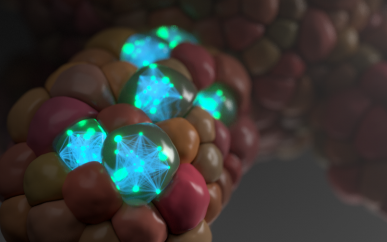
Artwork highlight signalling networks and relationships within organoid cells. Credit: Phospho Biomedical Animation.
Media contact
Henry Killworth
Tel: +44 (0) 207 679 5296
E: h.killworth[at]ucl.ac.uk