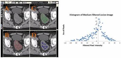
Heterogeneity is a key feature of malignancy associated with adverse tumour biology. Quantifying heterogeneity from imaging could provide a useful non-invasive biomarker. Heterogeneity on routinely acquired diagnostic images such as CT, MR, PET/CT, PET/MR, mammography, ultrasound can be quantified using texture analysis (TA) which may otherwise be imperceptible to the naked eye. One such TA-approach is the filtration-histogram technique where the filtration step extracts and enhances features of different sizes (comprising of fine, medium and coarse texture scales corresponding to 2-6mm in size) followed by quantification using histogram and statistical-based parameters. In our recent article [1] we describe the filtration-histogram technique of CTTA in more detail and demonstrated how these texture parameters relate to different aspects of heterogeneity such as number of objects, size, variation in density of the objects in relation to the background (parenchyma) of the tissue/lesion of interest.
As part of the qualification-process of CTTA as an imaging biomarker in oncology, a number of research-studies have demonstrated evidence towards:
* Biological correlates (hypoxia, angiogenesis, fibrosis, proliferation, genetic mutation) e.g. lung cancer [2, 3, 4]
* Technical validation (test-retest reproducibility and reliability assessment, multi-centre studies, degree of variability to different image acquisition parameters) e.g. lung cancer [5, 6]
* Clinical applications (e.g. lesion-characterisation, prognosis, treatment-response and prediction. e.g. lung-cancer [6, 7]
* Cost-effectiveness & clinical-utility (e.g. PET/CT lung MDT - clinical-adoption audit study); e.g. lung-cancer [8, 9]

CTTA has demonstrated potential clinical application as an adjunct to radiological and oncological work-flow in a number of cancer sites including lung, colorectal (primary & metastases), oesophageal, breast, prostate, head & neck, lymphoma, renal (primary & metastases), metastatic melanoma, soft-tissue sarcoma, gliomas, neuro-endocrine tumours etc. CTTA has demonstrated added-value in comparison and in combination (multi-parametric) with known existing biomarkers (size, attenuation, perfusion, metabolic activity, clinical stage and other clinical, blood markers and patient demographics) in several oncological applications. Recent applications in multi-parametric MR include identifying significant prostatic cancers within transition zone and amongst PIRADS 3 patients having significant cancers within the peripheral zone. Additionally, in rectal and breast cancer MRTA has demonstrated treatment response to chemo- and radiotherapy.
References:
References 1. Miles KA, Ganeshan B, Hayball MP. CT texture analysis using the filtration-histogram method: what do the measurements mean? Cancer Imaging. 2013 Sep 23;13(3):400-6. doi: 10.1102/1470-7330.2013.9045. Review. http://www.ncbi.nlm.nih.gov/pubmed/24061266
2. Weiss GJ, Ganeshan B, Miles KA, Campbell DH, Cheung PY, Frank S, Korn RL. Noninvasive image texture analysis differentiates K-ras mutation from pan-wildtype NSCLC and is prognostic. PLoS One. 2014 Jul 2;9(7):e100244. doi: 10.1371/journal.pone.0100244. eCollection 2014. http://www.ncbi.nlm.nih.gov/pubmed/24987838
3. Ganeshan B, Goh V, Mandeville HC, Ng QS, Hoskin PJ, Miles KA. Non-small cell lung cancer: histopathologic correlates for texture parameters at CT. Radiology. 2013 Jan;266(1):326-36. doi: 10.1148/radiol.12112428. Epub 2012 Nov 20. http://www.ncbi.nlm.nih.gov/pubmed/23169792
4. Ganeshan B, Abaleke S, Young RC, Chatwin CR, Miles KA.Texture analysis of non-small cell lung cancer on unenhanced computed tomography: initial evidence for a relationship with tumour glucose metabolism and stage. Cancer Imaging. 2010 Jul 6;10:137-43. doi: 10.1102/1470-7330.2010.0021. http://www.ncbi.nlm.nih.gov/pubmed/20605762
5. Chen SH*, Ganeshan B*, Fraioli F. Reproducibility of CT texture parameters by leveraging publicly available patient imaging datasets. British Journal of Radiology 2016 [under-review]
6. Win T, Miles KA, Janes SM, Ganeshan B, Shastry M, Endozo R, Meagher M, Shortman RI, Wan S, Kayani I, Ell PJ, Groves AM. Tumor heterogeneity and permeability as measured on the CT component of PET/CT predict survival in patients with non-small cell lung cancer. Clin Cancer Res. 2013 Jul 1;19(13):3591-9. doi: 10.1158/1078-0432.CCR-12-1307. Epub 2013 May 9. http://www.ncbi.nlm.nih.gov/pubmed/23659970
7. Ganeshan B, Panayiotou E, Burnand K, Dizdarevic S, Miles K. Tumour heterogeneity in non-small cell lung carcinoma assessed by CT texture analysis: a potential marker of survival. Eur Radiol. 2012 Apr;22(4):796-802. doi: 10.1007/s00330-011-2319-8. Epub 2011 Nov 17. http://www.ncbi.nlm.nih.gov/pubmed/22086561
8. Miles KA, Ganeshan B. Selection of patients for advanced non-small cell lung cancer for chemotherapy: Potential cost-effectiveness of CT Texture Analysis. In European Society of Radiology 2012, Vienna, Austria.
9. Miles KA. How to use CT texture analysis for prognostication of non-small cell lung cancer. Cancer Imaging. 2016 Apr 11;16(1):10. doi: 10.1186/s40644-016-0065-5. Review. http://www.ncbi.nlm.nih.gov/pubmed/27066905
 Close
Close

