The quantitative accuracy of PET and SPECT is affected by a phenomenon known as the partial volume effect (PVE). These effects are caused by the limited resolution of the scanner result in blurred images. Most partial volume correction (PVC) techniques utilise anatomical information from other imaging systems such as MRI or CT (see [1] for review). By combining information from the high resolution MRI or CT with the lower resolution PET or SPECT, PVEs can be reduced or removed, resulting in better estimation of the tracer distribution. With the availability of simultaneous PET/MR scanners, the application of PVC, particularly for brain imaging, is now much more feasible [2].
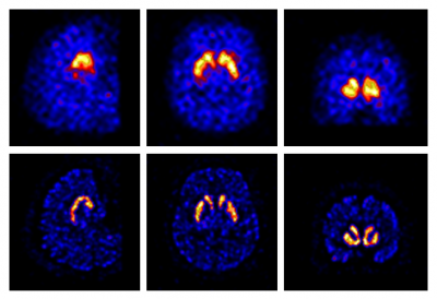
At the INM, we have developed a range of PVC algorithms for PET and SPECT applications. One PVC method, developed specifically for SPECT, is incorporated into an iterative reconstruction algorithm [3]. Fig. 1 shows an example with the DATSCAN tracer, used in the study of Parkinson's disease. We also developed the region-based voxel-wise (RBV) correction [4], which was applied in dementia studies with PET tracers that can image amyloid plaques (a hallmark of Alzheimer's disease). An alternative method is the iterative Yang (iY) technique [1], which is conceptually similar to RBV, but faster and simpler. Gaussian mixture model based deconvolution (GMD) [5] performs PVC while also maintaining the contrast in areas of the PET which may not be observable in MRI, such as hypometabolism in epilepsy. A method called Single Target Correction (STC) was also developed for the situation when only one image region needs to be corrected [6]. STC operates on a voxel-basis and does not require definition of any background regions.
It is important to be able to compare different PVC approaches under realistic conditions [7]. We are therefore currently in the process of developing a framework for the generation of test datasets based on actual patient data [8], which would also be made available to the wider nuclear medicine community. We have already made some of our software for PVC available as Open Source Software /nuclear-medicine/research/open-source-software

Figure 1: A brain SPECT study on a healthy volunteer using the tracer DATSCAN without (top) and with PVC (bottom), including sagittal (left), transaxial (middle) and coronal (right) sections.
References
[1] Erlandsson K, et al. (2012) Phys. Med. Biol. 57:R119-R159.
[2] Erlandsson K, et al. (2016) PET Clin 11:161-177, http://dx.doi.org/10.1016/j.cpet.2015.09.002
[3] Erlandsson K, et al. (2011) Nucl. Instr. Meth. A: 648: S85-88.
[4] Thomas BA, et al. (2011) Eur J Nucl Med Mol Imaging, 38:1104-19.
[5] Bousse A, et al. (2012) Phys. Med. Biol., 57:6681-6705.
[6] Erlandsson K, Hutton BF, J Nucl Med, 55 (Supplement 1):2123, 2014.
[7] Hutton BF, et al. (2013) Nucl. Instr. Meth. A, 702:29-33.
 Close
Close

