 Close
Close

The aim of the INSERT* project is to create a new multi-modality imaging tool for simultaneous SPECT and MRI studies by the development of a custom SPECT insert for commercially available MRI scanners. The INSERT project is funded by the EC under the FP7-HEALTH programme, and involves a number of collaborating groups throughout Europe (see http://www.insert-project.eu/). The system will be designed primarily for the study of brain tumours, mainly gliomas. The fundamental unit of the INSERT system is a compact gamma camera with high intrinsic resolution [1]. The principal task for our group is the detector-system and collimator design.
Whereas conventional SPECT systems consist of large rotating detectors, the INSERT system needs to fit inside the restricted space of the MRI bore. Also, the system should be stationary, as any detector motion would add complexity to the design and could interfere with the MRI signal. In relation to collimator design, a trade-off is always needed between sensitivity, resolution and FOV. For the current application, high sensitivity was considered more important than good spatial resolution.
The system optimisation is based on analytical calculations of sensitivity and resolution, as well as simulation and reconstruction of data corresponding to various geometric and anthropomorphic phantoms. For this purpose we developed a novel projection algorithm based on angular blurring [2]. We have investigated the use of both multi-pinhole and multi-slit-slat collimators [3]-[6]. A sensitivity similar to or higher than the standard collimator could be obtained with both multi-pinhole and multi-slit-slat systems. The former gives better uniformity and trans-axial resolution, while the latter gives better axial resolution.
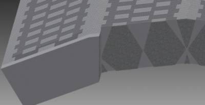
Figure 1: Reconstructions from simulated data for a multi-pinhole collimator.
We propose a new type of slit-slat collimation for the INSERT system: the mini-slit-slat (MSS) collimator [7] (Fig. 1). In contrast to a standard slit-slat collimator, in which the slit is continuous along the collimator, we explore the use of mini-slits to obtain improved angular sampling. In addition, we introduce the concept of interior slits, in which the slit aperture is located within the slat component, which thereby extend beyond the slit plane. If the resolution is fixed in both directions, the slat spacing can be increased, resulting in a sensitivity gain. The main advantage is allowing for minification without compromising the slat length and maximising the use of the space to optimise sensitivity.
Technical aspects of the system design are being addressed, such as collimator type [7], ring configuration [8], shielding [9], septal penetration, and geometrical calibration, and installation (Fig. 1). We have been using GATE to assess system performance. We are also currently performing experiments to compare mulit-pinhole and multi-slit-slat prototype collimators using a single detector module.
References
[1] Busca P, et al. (2014) Nucl. Instr. Meth. Phys. Res. A, 734:141-6.
[2] Bousse A, et al. (2013) Conf. Proc., Fully 3D reconstruction meeting.
[3] Erlandsson K, et al. (2013) Conf. Record,2013 IEEE Medical Imaging Conf.
[4]
Salvado D, Erlandsson K, Bousse A, Occhipinti M, Busca P, Fiorini C,
Hutton BF, "Collimator design for a clinical brain SPECT/MRI insert",
Eur J Nucl Med Mol Imaging: Physics 1 (Suppl 1): A21, 2014.
[5]
Erlandsson K, Busca P, Bukki T, Gola A, Salvado D, Butt A, Nemeth G,
Piemonte C, Hutton B, Fiorini C, "Design considerations for INSERT: A
new multi-modality SPECT/MRI system for preclinical and clinical
imaging", Journal of Nuclear Medicine 55, 2014.
[6] Salvado D,
Erlandsson K, Bousse A, Occhipinti M, Busca P, Fiorini C, Hutton BF,
"Collimator Design for a Brain SPECT/MRI Insert", IEEE Transactions on
Nuclear Science 62: 1716-1724, 2015.
[7] Salvado D, Erlandsson K,
Bousse A, van Mullekom P, Hutton BF, "Novel Collimation for SPECT/MRI",
IEEE Medical Imaging Conference, November 2014, Seattle, WA, USA.
[8]
Erlandsson K, Salvado D, Bousse A, Hutton BF, "Evaluation of a Partial
Ring Design for the INSERT SPECT/MRI System", Eur J Nucl Med Mol
Imaging: Physics 2 (Suppl 1): A47, 2015.
[9] Salvado D,
Erlandsson K, Hutton BF, "Shielding Requirements of a SPECT Insert for
Installation in a PET/MRI System", IEEE Medical Imaging Conference,
November 2015, San Diego, CA, USA.
* INtegrated SPECT/MRI for Enhanced Stratification in Radio-chemoTherapy
Figure 2: Multi-slit-slat collimator with 4 different phantoms (a uniform cylinder, a Derenzo phantom with rod-sizes of 6-11 mm, a Defrise phantom with parallel planes, and the Zubal brain phantom).