Using PET/CT and PET/MR we are working towards a better understanding of neurological conditions such as neurodegenerative disorders, neuro-oncology and epilepsy. The various metrics that we can derive from CT, and MR in particular, are being used together with dynamic PET data to produce multiple quantitative functional maps of dementia. Multi-modal techniques are also been used to characterize tumour metabolism and flow in the brain, and to devise novel methods of assessing epileptic foci.
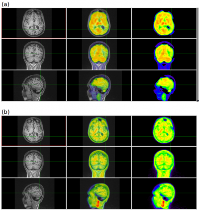
Figure 1:
PET/MR study using 1 injection of F18 Amyvid to display multiple
parameters (a) R1 images defined from dynamic PET show areas of
hypo-perfusion, some of which is caused by cortical atrophy (b)
Amyloid Imaging shows no build-up of amyloid plaques in the area of
hypo-perfusion.

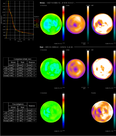
In cardiac PET, we continue to refine methodologies and better understand metrics such as coronary flow reserve and myocardial blood flow. Working on how these metrics fit clinically with other anatomical techniques such as CT coronary angiography and CT calcium score is a further area we are exploring. Beyond coronary artery disease, we are also looking at providing techniques and multi-parametric measures to better understand systemic diseases with cardiac findings such as sarcoidosis and amyloidosis.
Figure 2: An example of quantitative myocardial blood flow.

Our experience in kinetic modeling and dynamic PET is now being extended into oncological applications with the aim of maximizing the potential of readily available tracers such as FDG, fluorocholine and Rubidium-82. Using these tracers together with functional CT and MR imaging, we are working towards a better understanding of tumour physiology by relating our findings to histology.
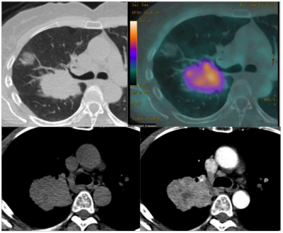
Figure 3:
Non small cell lung cancer shown on (a) a standard CT (b) a PET/CT
image with colour overlay signifying blood flow as measured by Rb82
PET, (c) a high resolution CT pre contrast and (d) post contrast.

 Close
Close

