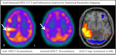
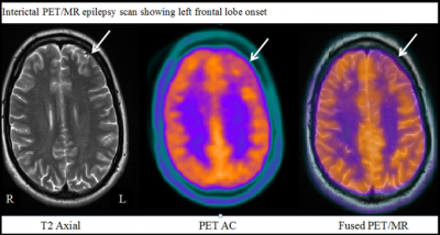
At INM a variety of imaging techniques are being used for the pre-surgical evaluation of drug-resistant epilepsy aiming to identify the seizure onset zone, in collaboration with The National Hospital for Neurology and Neurosurgery (NHNN).
We use FDG PET scans to map brain metabolism and identify hypometabolic areas which can point to the location of seizure onset. Voxel-based analysis comparing to an age-matched database of normal scans is in use to help pin-point the areas of significant hypometabolism.
Ictal/Interictal HMPAO SPECT scans are also performed to visualize brain perfusion patterns. My research focuses on SPECT/CT imaging using Ictal/Interictal subtraction analysis by Statistical Parametric Mapping. This analysis serves to highlight areas of significant hyperperfusion which are being used by researchers at NHNN to help plan intra-cranial EEG implantation.

Although PET/CT is the current clinical standard, the use of PET/MRI in epilepsy is also being explored at INM. Combining the excellent soft tissue contrast of anatomical brain MRI with metabolic information derived from PET can facilitate the seizure onset zone localization. Colleagues from INM (John Dickson) and UCL (Ninon Burgos) are working on the difficult task of MR-based attenuation correction for PET, which is a pre-requisite for quantitative PET analysis. Finally, new MR imaging techniques such as Arterial Spin Labelling and MR Spectroscopy are put to the test (Anna Barnes and Fransesco Fraioli) aiming to obtain complementary information about brain perfusion and tissue metabolites to improve seizure onset localisation.

 Close
Close

