The D-SPECT scanner (Spectrum Dynamics Medical, Caesarea, Israel) is based on solid state CZT-detectors which have better energy resolution as compared to traditional NaI(Tl)-based cameras [1]. Also the compactness of the CZT-detectors allow for faster scanning and dynamic studies. We are involved in various projects using this scanner.
Dynamic imaging
Dynamic cardiac SPECT can provide absolute quantitative values of myocardial perfusion, as opposed to semi-quantitative values obtained with static studies. The D-SPECT system is capable of performing dynamic studies, but due to the unconventional scanning procedure, there can be mismatch in the timing of the data acquisition and time-related effects, such as tracer kinetics and respiratory motion. We have investigated these effects using simulations [2].
Simultaneous dual radionuclide imaging
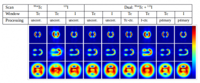
Simultaneous dual radionuclide SPECT studies can be useful in many situations. Advantages include automatically registered data as well as fewer scans for the patient. However, cross-talk and scatter correction is necessary in order to obtain quantitative images. We previously developed a correction algorithm for dual 99mTc and 201Tl imaging [3], which can be useful for simultaneous rest and stress cardiac perfusion studies. We have now developed correction algorithm for simultaneous 99mTc and 123I studies, based on a different formalism [4] (see fig. 1). This could be useful for studies of heart failure with 123I-MIBG or fatty acid metabolism with 123I-IPPA together with cardiac perfusion studies with 99mTc-MIBI, as a perfusion defect could lead to misinterpretation of the 123I data.

Figure
1: Results from 3 studies with a cardiac phantom containing 99mTc
only, 123I only and both radionuclides, respectively.
Short- and long-axis sections and bulls-eye plots are shown.
(X-ctr.=contributions from X.)
Angiogenesis imaging
We are involved in a trial aimed at investigating angiogenesis using a new radiotracer peptide (99mTc- Maraciclatide) in heart failure patients undergoing G-CSF and autologous bone marrow progenitor cells infusion. Maraciclatide is targeted at the αvβ3 integrins that is expressed by vessels undergoing angiogenesis. Patients included in the study undergo two baseline Maraciclatide scans at UCL, including a dynamic D-SPECT scan. The scans are repeated post-treatment, in order to establish whether Maraciclatide can be effective in detecting angiogenesis as part of a cell therapy approach. Preliminary results are encouraging.
References
[1] Erlandson K, et al. (2009) Phys. Med. Biol., 54:2635-2649.
[2] Costa F, et al. (2014) submitted to J Nucl Cardiol.
[2] Kacperski K, et al. (2011) Phys. Med. Biol. 56(5):1397-414.
[3] Holstensson M, et al. (2014), Phys. Med. Biol, under review.
 Close
Close

