 Close
Close

The Institute of Nuclear Medicine has extensive experience in SPECT and SPECT/CT imaging, with the department housing a number of state of the art hybrid SPECT/CT scanners. Combined
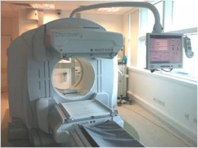
GE Discovery NM/CT 670 SPECT/CT camera
SPECT/CT
is an integrated imaging system which offers a combination of functional
data from a multi-head SPECT gamma camera and high- resolution
anatomical details from a multi-slice diagnostic CT scanner. The SPECT
functional data when fused with CT anatomical data provides the
clinicians with better visualisation of the images and enables them to
accurately define the exact location and extent of the disease. The
addition of anatomic information increases the sensitivity as well as
the specificity of imaging findings. The CT data is also used for
attenuation correction of the SPECT data, giving improved image quality.
The Institute of Nuclear Medicine performs a number of SPECT and SPECT/CT imaging procedures. The modality has already shown its benefits using different radio-labelled tracers in clinical practice and in research. Some of the indications where SPECT/CT has found to be of great benefit are as follows:
• Bone imaging
• Oncology imaging
• Cardiac imaging
• Neuro-imaging
• Endocrine imaging
• Sentinel node mapping