UCL Brain Sciences researchers are the first to carry out brain imaging of focal cortical epileptic seizures in the awake brain
15 August 2017
A team of researchers, led by Professors Dimitri Kullmann (UCL Institute of Neurology) and Matteo Carandini (UCL Institute of Ophthalmology), has developed a method of imaging brain activity to discover how it spreads during a focal cortical epileptic seizure.
 They were also able to then relate their propagation to cortical connectivity.
Focal cortical epilepsy affects about 0.5% of the population and is often resistant to pharmacological treatment. Patients with this disease can have malformations or lesions in a region of the cerebral cortex, although often the cause is not established. Epileptic seizures arise from a discrete focus and then propagate to other areas, and generalise to a large portion of the brain. Developing treatments for this disease requires understanding this propagation.
To study seizure propagation, Profs. Kullmann and Carandini, and Dr Rob Wykes, all from UCL Brain Sciences, joined forces to provide the first in vivo imaging of focal epilepsy in the awake cortex. Doctoral student Federico Rossi (Wellcome Trust PhD student in Neuroscience) who is hoping to take his viva in autumn) and Dr Wykes (Epilepsy Research UK Fellow) developed a powerful combination of pharmacology, electrophysiology, and imaging, in the visual cortex that enabled them to track how activity propagates to other areas of the cerebral cortex, and compare propagation to the normal connectivity between these areas.
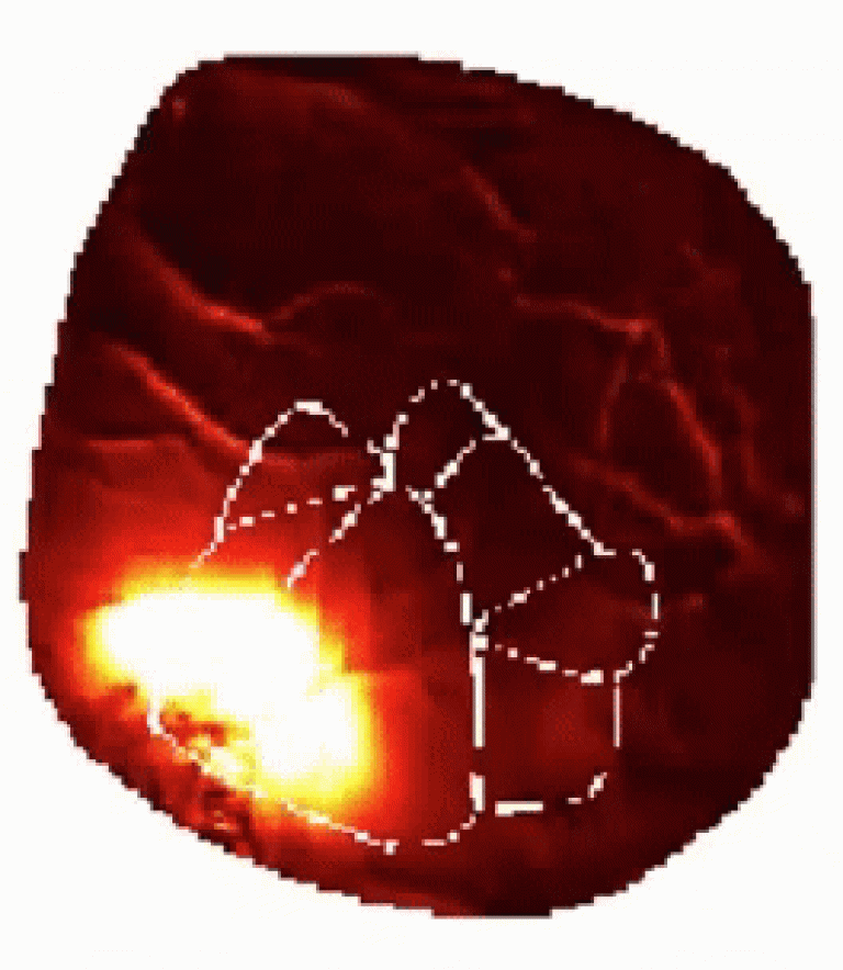
The images revealed that epileptic seizures first arose without leaving the focus but then propagated to other regions, and eventually generalized to large regions of cortex. Remarkably, even though seizures were hundred times stronger than normal activity, and likely involved a breakdown of synaptic inhibition, they propagated through the same pathways that channel activity during normal brain processing.
Prof. Carandini commented:
" I am delighted by the collaboration, and captivated by the compelling images of brain activity that we produced. These images provide the first description of how epileptic activity propagates from the focus in the awake cortex. We think they will be useful in guiding future work, both to address the mechanisms of propagation, and to test possible therapies."
This research was supported by the Wellcome Trust, Epilepsy Research UK and the Medical Research Council.
Image (gif)
The gif shows an example seizure recorded from almost one entire hemisphere of the cortex. The colourmap measures the intensity of neuronal activity (the brighter the colour the more intense the activity) and the white outline display the map of the organisation of the visual cortical areas.
 Close
Close

