Pigeons' homing skill not down to iron-rich beak cells
12 April 2012
The theory that pigeons' famous skill at navigation is down to iron-rich nerve cells in their beaks has been disproved by a new study published in Nature.

The study shows that iron-rich cells in the pigeon beak are in fact specialised
white blood cells, called macrophages. This finding, which shatters the
established dogma, puts the field back on course as the search for magnetic
cells continues.
"The mystery of how animals detect magnetic fields has just got more
mysterious" said Dr David Keays who led the study.
Dr Keays continued: "We had hoped to find magnetic nerve cells, but
unexpectedly we found thousands of macrophages, each filled with tiny balls of
iron."
Macrophages are a type of white blood cell that play a vital role in defending
against infection and re-cycling iron from red blood cells. They're
unlikely to be involved in magnetic sensing as they are not excitable cells and
cannot produce electrical signals which could be registered by neurons and
therefore influence the pigeon's behaviour.
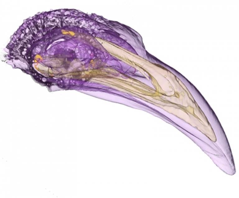
We employed state-of-the-art imaging techniques to visualise and map the location of iron-filled cells in the pigeon beak
Dr Mark Lythgoe
Dr Keays's lab, based at the Institute of Molecular Pathology in Vienna, worked
together with Dr Shaw from the University of Western Australia, and Drs Lythgoe
and Riegler from the UCL Centre for Advanced Biomedical Imaging in London.
"We employed state-of-the-art imaging techniques to visualise and map the
location of iron-filled cells in the pigeon beak" said Dr Mark Lythgoe.
The search for the actual mechanism that allows migratory birds, and many other
animals, to respond to the Earth's magnetic field and navigate around their
environment remains an intriguing puzzle to be solved.
"We have no idea how big the puzzle is or what the picture looks like, but
today we've been able to remove those pieces that just didn't fit," said Dr
Keays.
Image: State-of-the-art imaging at the UCL Centre for
Advanced Biomedical Imaging captured detailed maps of the pigeon beak. Magnetic
resonance imaging (MRI) revealed the external soft tissues (purple) and
micro-computed tomography (CT) exposed dense bony structures (yellow). (M.
Lythgoe, J. Riegler http://www.ucl.ac.uk/cabi/).
Media contact: Clare Ryan
The slideshow above captures the detailed map of the magnetic-free pigeon's beak that scientists used in the research.
Links:
Research in Nature
UCL Centre for Advanced Biomedical Imaging
 Close
Close

