Close
Close

Research from MSM@H is frequently disseminated in high-impact journals. See below our selected highlights and a full list of publication.
Publication Highlights
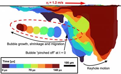
Keyhole fluctuation and pore formation mechanisms during laser powder bed fusion additive manufacturing
Huang, Y., Fleming, T.G., Clark, S.J., Marussi, S., Fezzaa, K., Thiyagalingam, J., ...Lee, P.D. (2022). NATURE COMMUNICATIONS, 13 (1), 1532-. doi:10.1038/s41467-022-28694-x
Abstract: Keyhole porosity is a key concern in laser powder-bed fusion (LPBF), potentially impacting component fatigue life. However, some keyhole porosity formation mechanisms, e.g., keyhole fluctuation, collapse and bubble growth and shrinkage, remain unclear. Using synchrotron X-ray imaging we reveal keyhole and bubble behaviour, quantifying their formation dynamics. The findings support the hypotheses that: (i) keyhole porosity can initiate not only in unstable, but also in the transition keyhole regimes created by high laser power-velocity conditions, causing fast radial keyhole fluctuations (2.5–10 kHz); (ii) transition regime collapse tends to occur part way up the rear-wall; and (iii) immediately after keyhole collapse, bubbles undergo rapid growth due to pressure equilibration, then shrink due to metal-vapour condensation. Concurrent with condensation, hydrogen diffusion into the bubble slows the shrinkage and stabilises the bubble size. The keyhole fluctuation and bubble evolution mechanisms revealed here may guide the development of control systems for minimising porosity.
The fatal trajectory of pulmonary COVID-19 is driven by lobular ischemia and fibrotic remodelling
Ackermann, M., Kamp, J. C., Werlein, C., Walsh, C. L., Stark, H., Prade, V., ... & Jonigk, D. D. (2022). EBIOMEDICINE, 85. doi:10.1016/j.ebiom.2022.104296
Abstract: Background: COVID-19 is characterized by a heterogeneous clinical presentation, ranging from mild symptoms to severe courses of disease. 9–20% of hospitalized patients with severe lung disease die from COVID-19 and a substantial number of survivors develop long-COVID. Our objective was to provide comprehensive insights into the pathophysiology of severe COVID-19 and to identify liquid biomarkers for disease severity and therapy response. Methods: We studied a total of 85 lungs (n = 31 COVID autopsy samples; n = 7 influenza A autopsy samples; n = 18 interstitial lung disease explants; n = 24 healthy controls) using the highest resolution Synchrotron radiation-based hierarchical phase-contrast tomography, scanning electron microscopy of microvascular corrosion casts, immunohistochemistry, matrix-assisted laser desorption ionization mass spectrometry imaging, and analysis of mRNA expression and biological pathways. Plasma samples from all disease groups were used for liquid biomarker determination using ELISA. The anatomic/molecular data were analyzed as a function of patients’ hospitalization time. Findings: The observed patchy/mosaic appearance of COVID-19 in conventional lung imaging resulted from microvascular occlusion and secondary lobular ischemia. The length of hospitalization was associated with increased intussusceptive angiogenesis. This was associated with enhanced angiogenic, and fibrotic gene expression demonstrated by molecular profiling and metabolomic analysis. Increased plasma fibrosis markers correlated with their pulmonary tissue transcript levels and predicted disease severity. Plasma analysis confirmed distinct fibrosis biomarkers (TSP2, GDF15, IGFBP7, Pro-C3) that predicted the fatal trajectory in COVID-19. Interpretation: Pulmonary severe COVID-19 is a consequence of secondary lobular microischemia and fibrotic remodelling, resulting in a distinctive form of fibrotic interstitial lung disease that contributes to long-COVID.
Correlative Synchrotron X-ray Imaging and Diffraction of Directed Energy Deposition Additive Manufacturing
Chen, Y., Clark, S. J., Collins, D. M., Marussi, S., Hunt, S. A., Fenech, D. M., ... & Lee, P. D. (2021). ACTA MATERIALIA, 209, 116777. doi:10.1016/j.actamat.2021.116777
Abstract: The governing mechanistic behaviour of Directed Energy Deposition Additive Manufacturing (DED-AM) is revealed by a combined in situ and operando synchrotron X-ray imaging and diffraction study of a nickel-base superalloy, IN718. Using a unique DED-AM process replicator, real-space imaging enables quantification of the melt-pool boundary and flow dynamics during solidification. This imaging knowledge was also used to inform precise diffraction measurements of temporally resolved microstructural phases during transformation and stress development with a spatial resolution of 100 µm. The diffraction quantified thermal gradient enabled a dendritic solidification microstructure to be predicted and coupled to the stress state. The fast cooling rate entirely suppressed the formation of secondary phases or recrystallisation in the solid-state. Upon solidification, the stresses rapidly increase to the yield strength during cooling. This insight, combined with the large solidification range of IN718 suggests that the accumulated plasticity exhausts the ductility of the alloy, causing liquation cracking. This study has revealed the mechanisms that govern the formation of highly non-equilibrium microstructures during DED-AM.
Imaging intact human organs with local resolution of cellular structures using hierarchical phase-contrast tomography
Walsh, C. L., Tafforeau, P., Wagner, W. L., Jafree, D. J., Bellier, A., Werlein, C., . . . Lee, P. D. (2021). NATURE METHODS, 18 (12), 1532-. doi:10.1038/s41592-021-01317-x
Abstract: Imaging intact human organs from the organ to the cellular scale in three dimensions is a goal of biomedical imaging. To meet this challenge, we developed hierarchical phase-contrast tomography (HiP-CT), an X-ray phase propagation technique using the European Synchrotron Radiation Facility (ESRF)’s Extremely Brilliant Source (EBS). The spatial coherence of the ESRF-EBS combined with our beamline equipment, sample preparation and scanning developments enabled us to perform non-destructive, three-dimensional (3D) scans with hierarchically increasing resolution at any location in whole human organs. We applied HiP-CT to image five intact human organ types: brain, lung, heart, kidney and spleen. HiP-CT provided a structural overview of each whole organ followed by multiple higher-resolution volumes of interest, capturing organotypic functional units and certain individual specialized cells within intact human organs. We demonstrate the potential applications of HiP-CT through quantification and morphometry of glomeruli in an intact human kidney and identification of regional changes in the tissue architecture in a lung from a deceased donor with coronavirus disease 2019 (COVID-19).

In-situ Synchrotron imaging of keyhole mode multi-layer laser powder bed fusion additive manufacturing
Chen, Y., Clark, S. J., Leung, C. L. A., Sinclair, L., Marussi, S., Olbinado, M. P., ... & Lee, P. D. (2020). APPLIED MATERIALS TODAY, 20, 100650. doi:10.1016/j.apmt.2020.100650
Abstract: The keyhole mode in laser powder bed fusion (LPBF) additive manufacturing can be associated with excessive porosity and spatter, however, the underlying physics in multilayer build conditions remain unclear. Here, we used ultra-fast synchrotron X-ray imaging to reveal this phenomena. We in investigated melt pool dynamics, keyhole porosity and spatter formation mechanisms and their impact in all layers of the build. We observed that the transient melt pool dynamics associated with the keyhole include: (I) keyhole initiation, (II) keyhole development, and (III) melt pool recovery. Porosity and spatter were associated with stages (II) and (III). We also discovered that droplet spatter can form due to the collapse of the keyhole recoil zone, causing molten particle agglomeration and ejection during stage (III). Our results clarify the transient dynamics behind the keyhole mode in a multi-layer LBPF process and can be used to guide the reduction in porosity and spatter in additive manufacturing.
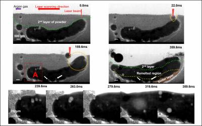
In situ X-ray imaging of defect and molten pool dynamics in laser additive manufacturing
Leung, C. L. A., Marussi, S., Atwood, R. C., Towrie, M., Withers, P. J., & Lee, P. D. (2018). NATURE COMMUNICATIONS, 9, ARTN 1355. doi:10.1038/s41467-018-03734-7
Abstract: The laser–matter interaction and solidification phenomena associated with laser additive manufacturing (LAM) remain unclear, slowing its process development and optimisation. Here, through in situ and operando high-speed synchrotron X-ray imaging, we reveal the underlying physical phenomena during the deposition of the first and second layer melt tracks. We show that the laser-induced gas/vapour jet promotes the formation of melt tracks and denuded zones via spattering (at a velocity of 1 m s−1). We also uncover mechanisms of pore migration by Marangoni-driven flow (recirculating at a velocity of 0.4 m s−1), pore dissolution and dispersion by laser re-melting. We develop a mechanism map for predicting the evolution of melt features, changes in melt track morphology from a continuous hemi-cylindrical track to disconnected beads with decreasing linear energy density and improved molten pool wetting with increasing laser power. Our results clarify aspects of the physics behind LAM, which are critical for its development.