We investigate atomic and nanoscale structures in semiconductors and two-dimensional (2D) materials using real and reciprocal space surface analytical techniques and simulation.
Schofield home ◊ People ◊ Research ◊ Publications ◊ Join us

Curiosity Driven Science with Impact
The ability to control matter at the fundamental length scale of individual atoms will enable the fabrication of entirely new classes of functional devices, from sensors to quantum computers. We conduct fundamental, exploratory reseach that is directed toward developing new understanding of matter at the atomic scale, as well as developing methods for atomic-scale assembly and construction.
Below you will find some examples of research from our group together with information about our laboratory. More detailed information on our published work can be found by accessing the papers in the Publications section of this web page.
- Atomic-scale quantum dots
Atomic-scale quantum dots in silicon

The hydrogen-terminated silicon (001) surface is a model system for exploring the properties of atomic-scale quantum dots, which can be fabricated by removing individual hydrogen atoms one at a time with the STM tip. The absence of a single hydrogen atom produces a "dangling bond", which can be considered as a deep-level atomic-point defect. Creating assemblies of closely-spaced dangling bonds leads to the formation of artificial molecular orbitals.
- Two-dimensional (2D) Materials
Charge Density Waves on the Graphene Sheets of the Superconductor CaC6
Systems of reduced dimensionality, e.g., two-dimensional (2D) crystals, can exhibit spontaneous reorganisation of their free charge density, forming what are often referred to as charge density waves (CDWs). CDWs are usually the result of electron-phonon coupling (i.e., coupling of the electrons with the vibrational modes of the crystal) or electron-electron coupling (i.e., coupling of the electrons with themselves). The signature of CDWs are a long range modulation of the charge density, which can be commensurate or incommensurate with crystal lattice, and the opening of an energy gap at the Fermi energy. The ability of the scanning tunnelling microscope to map the density of states with both atomic-scale spatial resolution and meV energy resolution makes it the ideal tool with which to explore CDWs in 2D materials.

- Mapping dopant wave functions in silicon
Imaging Donor and Acceptor States in Silicon
Atoms from group V of the periodic table, e.g., phosphorus, arsenic, or antimony, can substitute for a silicon atom in a crystal of silicon. Since silicon is a group IV element, this results in a localised positive charge of +1e at the impurity site. This charge can localise an electron from the conduction band and in doing so forms a hydrogenic state. The orbitals of this hydrogenic defect are analogous to those of the hydrogen atom with some perturbation due to the effects of the crystal lattice. Using the scanning tunnelling microscope we can image the probability densities of these long-ranged quantum states (see above) produce energy-resolved spatial maps of their density of states. Hydrogenic defect states are currently being explored for their potential use as quantum bits ("qubits") in various solid-state quantum computer architectures. The details understanding of their properties that can be gained from STM/STS measurements of individual defects and their interactions provides detailed information on these states that cannot be determined by any other method.

[Single dopants in semiconductors. Editorial Comment. Schofield, SR; Rogge, S]
- Organic functionalisation of semiconductor surfaces
Organic functionalisation of semiconductors
One of the focuses of the group is developing methods to incorporate molecular functionality with traditional semiconductor electronics. We approach this by depositing organic molecules onto the technologically ubiquitous (001) crystal face of silicon under ultrahigh vacuum. The functionalised surfaces we investigate using both scanning tunnelling microscopy (STM), and synchrotron-based photoelectron spectroscopy techniques, including x-ray photoelectron spectroscopy (XPS) and near edge x-ray adsorption find structure (NEXAFS) spectroscopy. We have performed synchrotron experiments at the Swiss Light Source, the Australian Synchrotron, the National Synchrotron Radiation Research Centre (NSCCR; Taiwan), and have collaborated with scientists from the Diamond Light Source (UK).
Methoxyacetophenone adsorption to Si(001)
We have investigated the coverage-dependent behavior of p-methoxyacetophenone adsorption to atomically-clean Si(001) using X-ray photoelectron spectroscopy and density functional theory calculations. This compound exhibits a high selectivity of adsorbate species formation by forming only two distinct adsorbate structures at low coverage, with a third configuration forming at high coverages. This coverage-dependent but well-defined behavior is promising in designing functional organic−inorganic interfaces on silicon. This work resulted in our second publication in our collaboration with the Teplyakov group at the University of Delaware.
[Dissociation of CH3-O as a Driving Force for Methoxyacetophenone Adsorption on Si(001), Journal of Physical Chemistry C, 2019]
- Low-temperature Scanning Tunnelling Microscope Laboratory
Scanning Tunnelling Microscopy
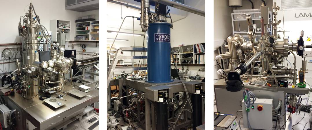
We have three ultrahigh vacuum (UHV) scanning tunnelling microscope (STM) systems that are run by two principal investigations, Steven Schofield and Neil Curson [Neil Curson's LCN web page].

- (Left): 4-Kelvin UHV-STM optimised for spatially resolved tunnelling spectroscopy and the creation of atomic-scale structures.
- (Middle): 6-Tesla vector magnet field, 1.5-Kelvin UHV STM system. Optimised for tunnelling spectroscopy of magnetic systems.
- (Right): Variable temperature UHV STM. This system is used solely for the fabrication of atomic-scale dopant structures in silicon.
Acknowledgements
Our research is funded by the Engineering and Physical Sciences Research Council (EPSRC). Additional support and collaborations are gratefully acknowledged below.

 Close
Close

