Visualising the brain's communications networks

14 December 2014
Brain surgeons are using open-source software to help them avoid damaging major pathways in the brain during neurosurgery, greatly reducing the risk of patients experiencing permanent cognitive damage, for example in visual, verbal or motor skills. The software - the Camino toolkit - is used in surgery for treating epilepsy and brain tumours.
The toolkit, developed by researchers at UCL Computer Science, enables neurosurgeons to visualise, before surgery, the white matter fibre pathways that form the brain's communication network. This helps them avoid cutting these fibres during the operation, helping patients avoid severe cognitive damage unrelated to the original problem that led to the surgery.
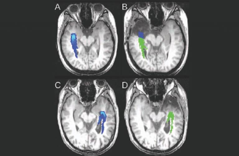
Surgeons use algorithms developed for Camino to identify pathways of white matter fibres in the brain from pre-operative MRI scans of patients with epilepsy, damage to which can lead to visual deficits that would, for example, prevent driving. At the National Hospital for Neurology and Neurosurgery in London, a specialised image analysis system is used to support doctors carrying out surgery on patients whose seizures are not controlled by standard medicines. They use the system on roughly one patient a week and early evaluations showed that the patients whose visual pathway was displayed to the surgeon via the Camino-based system had significantly better retention of visual skills than those operated on without the system.
This technique is a significant benefit to surgeons planning brain tumour resections, because they get a much clearer picture of white matter pathways near the tumour. Dr Alberto Bizzi, HUMANITAS Research Hospital, Milan
UCL's researchers continue to develop new imaging techniques to enhance clinicians' visualisation of the brain's networks. Since 2012, neurosurgeons in Milan planning interventions to remove brain tumours have replaced their existing connectivity mapping with one of the new methods, known as NODDI. It is less vulnerable to the effects of tumours, such as swelling, so shows the fibres near tumours much more clearly. As with the epilepsy patients, this helps surgeons avoid damaging the vital white matter pathways, and reduces the likelihood and severity of common cognitive deficits such as are verbal, sensory, or motor problems.
Funding from the EPSRC.
Image
- Pre-operative and post-operative images of two patients who underwent an anterior temporal lobe resection as treatment for severe epilepsy.
 Close
Close

