First prostate cancer patient scanned using hyperpolarised MRI in Europe
23 August 2017
A University College Hospital prostate cancer patient has undergone diagnosis using hyperpolarised Magnetic Resonance Imaging (MRI) as part of a UCL clinical trial. Led by Dr Shonit Punwani, the trial aims to develop more accurate and personalised treatment for cancer.

The scan was the first time this experimental imaging method was used in Europe to scan a patient with prostate cancer and was able to help doctors confirm the location and state of the patient’s tumour. The clinical trial is a novel programme hoping to revolutionise diagnosis, risk stratification and therapy for people with cancer, based on innovations in MRI technology.
The work builds on the results of the PROMIS study published in February, which showed that many prostate cancer patients were undergoing unnecessary biopsy procedures, which can cause side effects including pain, bleeding and infection. The study also indicated that multi-parametric magnetic resonance imaging used as a triage test can avoid unnecessary biopsies and improve diagnostic accuracy.
Improved diagnosis
It is hoped that by using hyperpolarised MRI and multi-parametric MRI together, clinicians can further improve diagnostic accuracy by giving a clearer picture of the exact tumour location. Furthermore, by understanding the metabolic activity, clinicians hope to differentiate between aggressive or non-aggressive tumours, helping to guide treatment decisions.
The clinical trial at UCL uses a method which sees the patient injected with a traceable sugar before undergoing an MRI scan. Before injection, the sugar is put through a process called hyperpolarisation using specialised equipment set up at UCLH. This boosts the signal produced by the sugar by more than 10,000 times. The metabolic conversion of the sugar in the location of tumour can be picked up by MRI to determine where the tumour is, and may be helpful in determining how aggressive the tumour is.

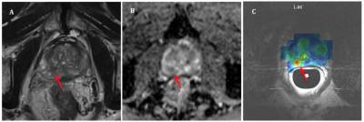
An axial T2 weighted MRI (A: top image) and Apparent Diffusion Coefficient map (B: middle image) acquired as part of a routine multi-parametric MRI study of the prostate. 13C spectroscopic lactate image (C: bottom image) demonstrates high levels of lactate at the position of the biopsy positive Gleason 3+4 tumour site (red arrow).
Dr Shonit Punwani, UCL Reader in Magnetic Resonance and Cancer Imaging (Centre for Medical Imaging) and UCLH honorary consultant radiologist, said: “The multi-disciplinary research team have spent the past two years installing and testing the equipment, creating standard operating procedures and planning the study in great detail. We are really pleased to have scanned our first patient and we will continue to work closely with other sites, in the UK and internationally, to develop this technology further.” Dr Punwani is the principal investigator of the Hyperpolarised MRI study which is developing the technology so that a full clinical evaluation can take place.
Prof. Mark Emberton, UCL Dean of the Faculty of Medical Sciences and UCLH honorary consultant urologist, said: “The technology offers a real opportunity to deal with the problem of overtreatment of patients with prostate cancer. Most prostate cancers will be non-aggressive and so being able to interrogate their metabolism may help patients with indolent disease avoid treatment.” Prof Emberton was the principal investigator on the PROMIS study.
Prof. David Lomas, Vice-Provost (Health) at University College London, added “I am proud of the UCL team for achieving this milestone. UCL was one of 3 UK institutions to receive Medical Research Council (MRC) funding in 2015 for the installation of the SpinLab Hyperpolariser. UCL has a strong history of collaborative working and this is reflected in the development of this technology”.
The study is supported by funding from the Medical Research Council, the Wellcome Trust, the National Institute of Health Research UCLH Biomedical Research Centre, Cancer Research UK, and philanthropic funding provided by the Mitchell’s Charitable Trust.
The Spinlab hyperpolariser from GE is situated at the University College Hospital Macmillan Cancer Centre and designed to work in conjunction with existing MRI equipment to provide non-invasive metabolic assessment of tumours. The traceable sugar injection was developed at the UCL Good Manufacturing Practice lab.
Further information
- Source: UCLH and UCL Institute of Healthcare Engineering
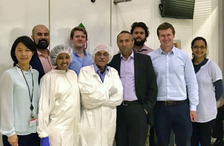
- Image top: from Left to Right: Fiona Gong (Project Manager), Arash Latifoltojar, Ramla Awais (GMP lab manager), William Devine, Hassan Jeraj (Quality Assurance Manager), Shonit Punwani (Principal Investigator), Edward Johnston, James O’Callaghan (MR Physicist), Mrishta Brizmohun.
- Professor Mark Emberton academic profile
- Dr Shonit Punwani academic profile
- UCLH
- NIHR University College London Hospitals Biomedical Research Centre
Related news
Launch of new clinical research facilities to advance cancer diagnostics and therapies at UCL/UCLH
The final stages of an £8.9 million award to advance cancer medical research facilities at UCL/UCLH, were celebrated last night with a launch event marking the implementation of new cancer genomics and imaging technologies that will support two world-class cancer research programmes.
 Close
Close


