PET/CT
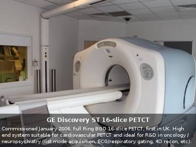 | 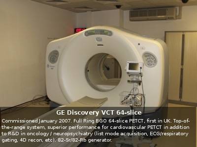 |
PET/MR
The Institute of Nuclear Medicine at UCH was the first department in the UK to set up a clinical PET/CT service to great success, currently imaging approximately 100 patients per week, has now taken on the exciting challenge of being the first to set up a clinical PET/MR. The latest development in hybrid imaging from Siemens in Germany started operation; on the 2 April this year. It has been established as an important part of the brand new UCH Macmillan Cancer Centre to provide its patients with an holistic experience where all essential facilities are under one rather spectacular roof.
But the scanner is not only used for imaging patients with cancer but is also used to investigate heart and brain function. MRI image of the brain with a radioactive sugar scan showing the fully functioning parts of the brain overlaid. We are looking sideways on at a slice right through the middle of the brain. This scanner combines positron emission tomography (PET) with magnetic resonance imaging (MRI). This is quite an engineering marvel as the electronics used by a traditional PET camera don't work inside a high magnetic field associated with MRI scanners. Instead Siemens redesigned the PET technology so that it could.
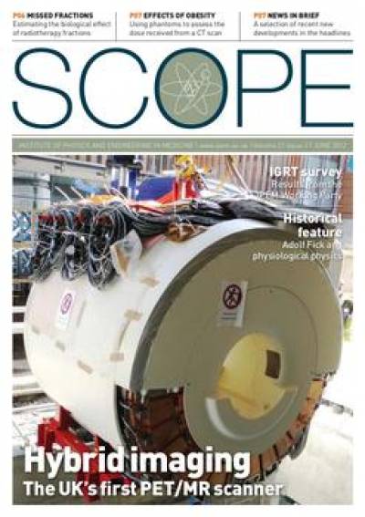 | 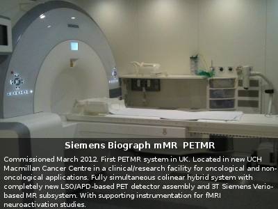 |
Basically this scanner looks like an MRI from the outside but the difference is that it also has a ring of PET detectors under the cover in the middle of the magnet. If your doctor requests this exam for you your experience will in fact be very similar to that of having an MRI scan but you will also have an injection of a very small amount of radioactive sugar before hand. The exam will take anywhere between 30 mins and half an hour but you can bring your own music or podcasts to listen to.
In order that the radioactive sugar goes to the right place you will not be able to eat anything for about 6hrs before you arrive and in order to avoid taking anything metal into the MRI scanning room you will be asked to change into a hospital gown or pyjamas.
Once the exam has finished you will be able to eat and go about your normal business although it is advised to keep your distance from small children and crowds for the rest of the day.
If you have any other queries please contact Dr Anna Barnes (anna.barnes@uclh.nhs.uk)
SPECT/CT
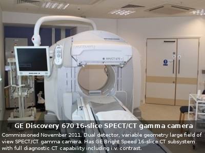
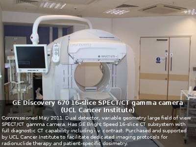
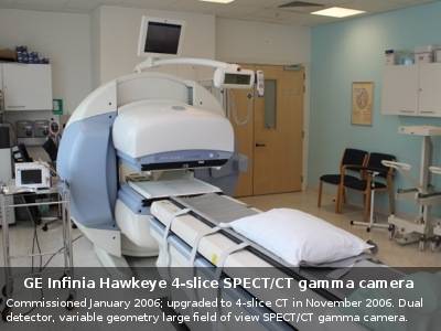
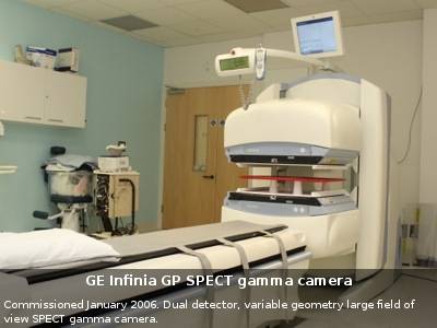
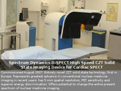
The Institute of Nuclear Medicine houses a number of gamma cameras capable of SPECT imaging. Following an injection of a radioactive tracer, a SPECT scan produces three-dimensional images of body function. A SPECT scan is commonly performed in combination with a CT scan, with the CT component providing anatomical information. Functional and anatomical information of the body can in this way be obtained in one single appointment. The fused data provides better visualisation of the images and enables the clinicians to accurately define the exact location and extent of the disease.
The Institute of Nuclear Medicine has extensive experience in a number of SPECT and SPECT/CT imaging procedures, both for routine and research purposes. Some of the indications where SPECT CT has found to be of great benefit are as follows:
• Bone imaging - The musculoskeletal system is imaged to investigate bone disease and pain
• Oncology imaging - Tumour sites are localised for both diagnostic and post-therapeutic purposes
• Cardiac imaging - Functional images of the heart are acquired, normally in conjunction with ECG monitoring of the cardiac cycle
• Neuroimaging - Images are acquired for a number of brain conditions, including movement disorders, dementia and epilepsy
• Endocrine imaging - Parathyroid adenomas are localised, commonly for surgical workup
• Sentinel node mapping - The lymphatic drainage from tumour sites is imaged
 |  |
 |  |
 |
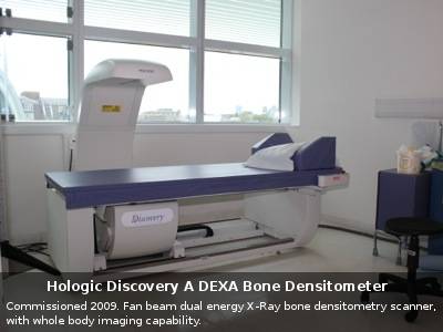
Bone Density
 |
 Close
Close

