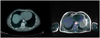
According to Cancer Research UK there is approximately 9,000 new case of oesophageal cancer in the UK every year (between 2013-2015) and is the 13th most common cancer in the UK. Approximately 12% of the people in England and Wales survive their disease for five years or more.
Accurate estimates of patient survival with oesophageal cancer can aid oncologists in making better treatment outcome decisions. The use of clinical information, pathological data, dynamic and molecular imaging can be used to assess to see whether they can provide prognostic information and personalised estimates of survival which may influence the treatment choices used.
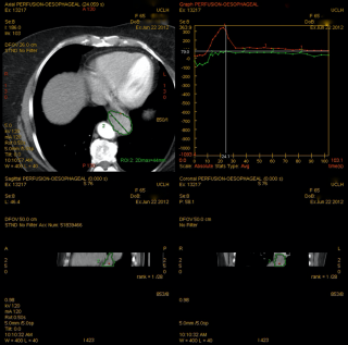
Both dynamic contrast-enhanced computed tomography and CT texture analysis can provide an invaluable non-invasive method of quantifying tumour heterogeneity and estimating tumour vascularization in-vivo and have the potential to provide information on tumour aggression and predict patient survival. In addition dynamic contrast-enhanced MRI can be used to assess vascular permeability and can potentially be used in follow up imaging of patients eliminating the use of radiation.
The objective of this single institution tertiary centre study is to evaluate whether a combination of pre-treatment dynamic contrast-enhanced computed tomography parameters (tumour volume, arterial flow, blood volume and permeability), tumour heterogeneity (with the use of CT texture analysis), metabolic uptake of the tumour on PET-CT imaging and metabolic markers in relation to hypoxia (with the use of histology staining patterns) can predict patient tumour aggression and prognosis of oesophageal or gastroesophageal junction cancer. This will be continued as a prospective study in combination with functional and anatomical imaging with the use of PET-MR.
Content by Dr Christine Tang
 Close
Close


