 Close
Close

Interstitial Lung Disease or ILD, is an umbrella term for many different lung conditions and is characterised by the progressive destruction of the interstitium (the tissue that surrounds the air sacs in the lung). Idiopathic Pulmonary Fibrosis or IPF for short, is the most severe form of the disease and has a poorer prognosis than many cancers with an average survival of just 3 years after diagnosis.
Although CT scans are essential for diagnosing Interstitial Lung Disease and can be used to produce a number of promising measures of the disease, such techniques only map the structural abnormalities of the lungs, with no consideration to the underlying inflammatory processes which are believed to be the dominant element of the disease.
At the Institute of Nuclear Medicine, we have been running a research project since 2006 aimed at correlating the severity of interstitial lung disease with the metabolic activity of lung cells (which reflects the degree of inflammation). While we don’t know the exact cellular mechanisms underlying the observed increase in FDG-PET signal (FDG is a radioactive tracer that is taken up by metabolically active cells), there is evidence that it is at least in part, caused by metabolically active cells in the lungs of ILD patients. In this way, FDG-PET has been shown to be a useful tool in improving our understanding of the pathogenesis and prognosis of Interstitial Lung Disease and we are currently exploring it’s potential to detect metabolic changes in the lungs before the damage becomes apparent on CT - which would be helpful in mild or early disease. Furthermore, we recently completed a prospective clinical trial involving IPF patients that revealed an important prognostic role for FDG/PET to track the progression of disease, and which may provide a useful measure of disease burden for use in future clinical studies looking to test new treatments.
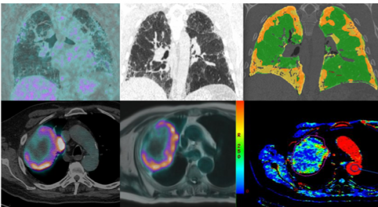
The top row images represent an example of the INM pathway for patients with idiopathic pulmonary fibrosis. We scan all patients with a combined protocol HRCT/PET-CT. this approach is twofold, allowing to obtain anatomical (HRCT) and functional (FDG PET)imageThe images at the bottom represent an example of multiparametric imaging in lung cancer. From right to left: FDG PET/CT, PET/MRI and perfusion map following dynamic injection of contrast media in CT.
Our centre has several collaborators both within the campus and externally within industry including: GSK, The Mayo Clinic and Imbio (with whom we are working with to develop a platform for CT quantification of ILD).
To date and to the best of our knowledge we are the first in world to combine a protocol of High Resolution CT and FDG-PET in a single exam and apply a computed algorithm for the CT quantification of Interstitial Lung Disease.
Content by Dr Francesco Fraioli.