The tradeoff between field of view (magnification) and spatial resolution (numerical aperture) in a conventional optical microscope limits its capacity to capture information.
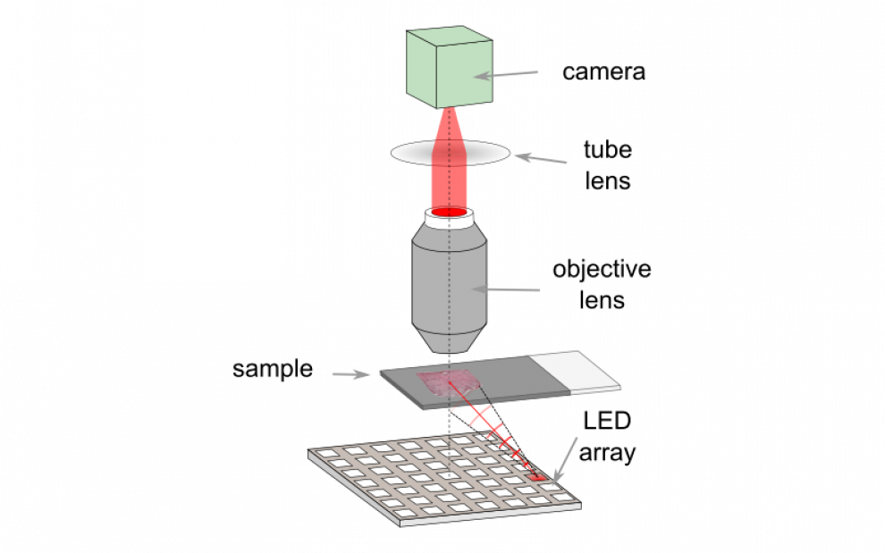
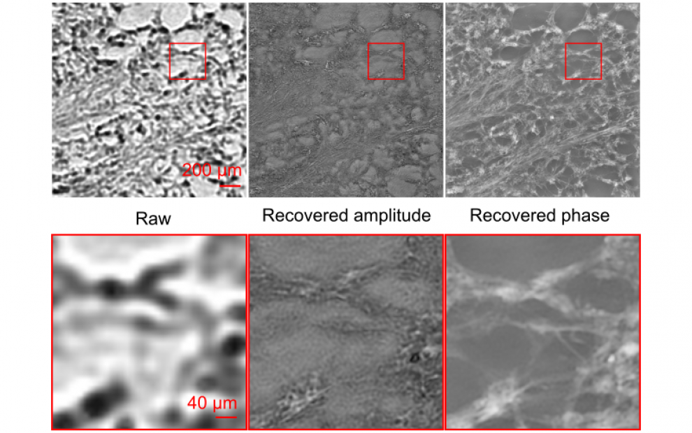
This is particularly problematic for biological and biomedical imaging applications, which often require high resolution images including large cell populations or large tissue areas in order to make reliable, statistically robust inferences. Fourier Ptychographic Microscopy (FPM)1 offers a way to combine low magnification and high resolution by illuminating the sample off axis using an LED array. Images captured under illumination at different angles contain information from different regions of the underlying object spectrum. Combining the information contained within the image sequence using iterative phase retrieval allows recovery of a complex image (amplitude and phase) with significant higher spatial resolution.

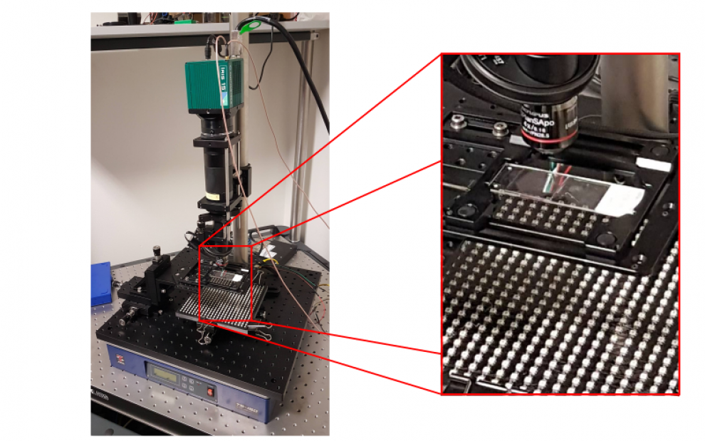
In the TouchLab we have built a low-cost FPM system using off-the-shelf components, a commercially available LED matrix and a microcontroller. We have been exploring methods to use FPM to visualize thicker samples2 and reduce the effects of background and image noise on the quality of reconstructed images3.

We are working in collaboration with clinicians and other researchers to apply FPM for a range of diagnostic imaging applications including the detection of malarial parasites in peripheral blood films and the identification of tumour margins in histological tissue sections.

1https://www.nature.com/articles/nphoton.2013.187
2https://doi.org/10.1364/BOE.11.000215
3https://doi.org/10.1364/OE.403780
 Close
Close

