Fluorescence
Optical microscopy has been wonderfully effective at elucidating many aspects of cell structure and behaviour on a tiny scale, but often details cannot be distinguished under ordinary 'brightfield' illumination. To more clearly identify particular features or processes, we can instead attach fluorescent markers to them that emit light of one colour band when illuminated with a different one. By filtering out all the light in the illumination range, we are left with only the emission light, and hence only the details of interest.
There are numerous methods for attaching markers to different cell regions, depending very much on what it is one wants to identify. Many derive from the availability of fluorescent proteins that, given suitable encouragement, the cell can manufacture for itself alongside the proteins that participate in the target function. Other techniques depend on particular chemical properties of the fluorescent molecule used.
In this project we are currently using two kinds of fluorescent tag.
Styryl dyes such as FM 1-43 and FM 4-64 are amphipathic molecules that readily enter and leave the plasma membrane but are prevented by their permanent charge from passively diffusing across it. They are barely fluorescent in water, but fluoresce strongly when partitioned into the hydrophobic environment of the membrane. They can thus be used as markers of vesicular activity. The membrane is first stained with the dye and then, after a short delay, washed out. Patches of membrane that have been endocytosed in the interim retain their fluorescence, and these internalised puncta can be taken to identify presynaptic release sites.
GAD65-GFP transgenic mice express green fluorescent protein (GFP) under the control of the promoter for an enzyme that catalyses the production of the inhibitory neurotransmitter γ-aminobutyric acid (GABA). This leads to selective GFP expression in inhibitory neurons, and specifically the cerebellar interneurons we are interested in. The resulting fluorescence is bright, stable and distinct.
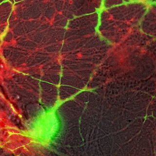
FM 1-43 does not combine well with GFP because the two emission spectra overlap considerably, but FM 4-64 and GFP are well separated, showing virtually no crosstalk. We therefore use these together to help target our recordings. The image above is an example. The red areas are those stained by FM 4-64, while the green areas are GFP-expressing interneurons. The greyscale background is what we see just with plain white illumination.