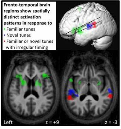
Magnetic resonance imaging (MRI) is most often used as a clinical tool to provide images of the brain structure. However, functional MRI (fMRI) provides images of brain function. This technique measures fluctuations of the oxygen concentration within the blood. As a region of the brain becomes more active, it requires more oxygen to perform. The conventional method of using fMRI is to provide the user a task (such as visual identification, decision-making, memory tests) while they are in the scanner. The fMRI images can then be used to determine which part of the brain "lights up" in relation to the task (as seen in the picture). The fMRI contrast results in the picture have been plotted on group mean structural brain scan images and show the blood oxygen level-dependent (BOLD) responses to different types of auditory stimuli for a group of older healthy controls (Agustus & Warren, 2012).
Another method of fMRI that is increasingly popular in dementia research is "resting state" fMRI. In this form of fMRI, the participant is not asked to perform a task but rather lie down in the scanner. The natural brain activity is then recorded. Three primary "resting state networks" have been identified in association with this activity: the default mode network (DMN), the executive network, and the salience network. These networks consist of connected nodes (brain regions) of activity that fire in sequence with each other. Research in Alzheimer's disease (AD) indicates that connectivity between the nodes of such large-scale neural networks weakens in AD. At the same time, there is also evidence of increased activity in AD relative to normal controls between some of the network nodes, suggesting that hyperactivity of these areas may be a sign of the disease.
 Close
Close


