 Close
Close

Photoacoustic imaging is a new biomedical imaging modality based on the use of laser-generated ultrasound. It is widely viewed as one of the most exciting and promising imaging techniques to have emerged in recent years. The technique relies upon the absorption of low energy nanosecond pulses of visible or near infrared laser light by specific tissue chromophores to excite broadband ultrasound waves. These waves are encoded with the optical properties of the tissue and, by recording them over the tissue surface using an array of ultrasound receivers, a 3D absorption based image can be reconstructed. The fundamental advantage of the technique is that, by encoding optical absorption on to acoustic waves it avoids the penetration depth/spatial resolution limitations of purely optical imaging techniques such as light microscopy or diffuse optical tomography that arise from the strong optical scattering of tissue. At the same time it retains the high molecular based contrast and spectralspecificity of optical methods enabling visualisation of anatomical features indistinguishable with other imaging modalities such as ultrasound.
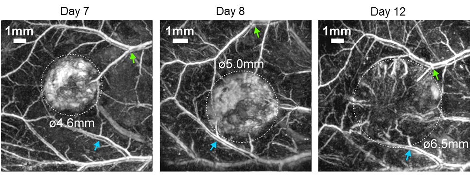
Figure 1
Longitudinal photoacoustic image of tumour vasculature development in a mouse model of colorectal cancer.
Laufer J, et al (2012) In vivo preclinical photoacoustic imaging of tumor vasculature development and therapy, Journal of Biomedical Optics 17(5), 056016
CABI has a novel high resolution preclinical photoacoustic scanner, based upon on a highly sensitive optical ultrasound detector developed in the Department of Medical Physics and Biomedical Engineering at UCL. The high sensitivity of the detector combined with novel image reconstruction algorithms enables the acquisition of 3D non-invasive in vivo images at depths approaching 1cm with sub 100µm resolution, making the scanner a powerful preclinical imaging tool. Using the scanner, the strong optical absorption of endogenous haemoglobin in tissue has been exploited for studying processes characterised by changes in the structure and function of the vasculature. Examples of such studies include investigating tumour vascular development in whole subcutaneous tumours (see figure 1) as well as the response of tumours to vascular targeted therapy, in a manner not previously possible with existing imaging modalities. Other studies that have been undertaken include investigations into the vascularisation of tissue engineered scaffolds, changes to vascular architecture in mouse models of kidney disease and embryo development. In addition, exogenous contrast, in the form of targeted contrast agents or genetically expressed absorbers have been used to study processes at a molecular level. An example is the in vivo imaging of genetically encoded contrast in order to visualise cell populations (see figure 2).
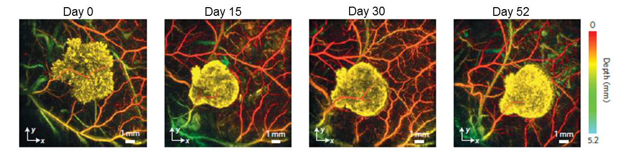
Figure 2
Photoacoustic images of cells genetically engineered to express tyrosinase, an enzyme which catalyses the formation of the pigment eumelanin
Jathoul AP, et al (2015) Deep in vivo photoacoustic imaging of mammalian tissues using a tyrosinase-based genetic reporter, Nature Photonics 9, 239-246
For information about Photoacoustics please have a look at the webpage of the UCL Photoacoustic Imaging Group
See the collaborations page for information on access to this system