Old world monkey had tiny, complex brain
3 July 2015
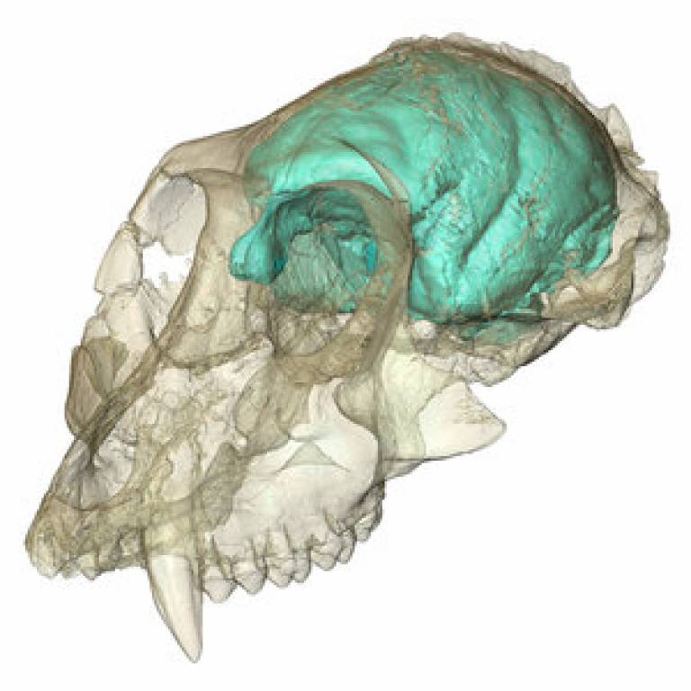
The brain of a 15 million year old monkey has been visualized for the first time by a team led by Professor Fred Spoor (UCL Cell & Developmental Biology).
 The 3D
computer model shows that the brain is much smaller and has more folds than
expected, supporting the idea that brain complexity can evolve before brain
size in the primate family tree.
The ancient 'Old World' monkey is known scientifically as Victoriapithecus and first made headlines in 1997 when its fossilised skull was discovered on an island in Kenya's Lake Victoria, where it lived 15 million years ago. As fossils from this era are rare, the skull was one of the only clues to the early brain evolution of Old World monkeys. Before it was studied, scientists didn't know if primate brains got bigger first and then more complex, or vice versa.
The findings, published today in Nature Communications by UCL, Duke University, Max Planck Institute for Evolutionary Anthropology and New Mexico State University, offer new clues for how primate brains changed over time.
"In the part of the primate family tree that includes apes and humans, the thinking is that brains got bigger and then they get more folded and complex," co-author Lauren Gonzales (Duke University) said. "But this study is some of the hardest proof that in monkeys, the order of events was reversed - complexity came first and bigger brains came later."
The findings also lend support to claims that the small brain of the extinct human relative Homo floresiensis, whose 18,000-year-old skull was discovered on a remote Indonesian island in 2003, isn't as remarkable as it might seem. In spite of its tiny brain, Homo floresiensis was able to make fire and use stone tools to kill and butcher large animals.
This study shows the power of modern digital imaging methods to get essential information out of precious fossils that would otherwise remain unknown.
Professor Fred Spoor
The team used high-resolution X-ray imaging to peer inside Victoriapithecus' cranial cavity to create a three-dimensional computer model of what the animal's brain likely looked like. The micro-CT scans of the creature's skull show that it had a tiny brain, at 36 cubic centimetres, relative to its body. This is less than half the volume of monkeys of the same body size living today.
Co-author Professor Spoor (UCL & Max Planck Institute for Evolutionary Anthropology), who is an expert in studying fossils with computed tomography and 3D visualization, said: "This study shows the power of modern digital imaging methods to get essential information out of precious fossils that would otherwise remain unknown. The CT scans revealed numerous distinctive wrinkles and folds of the brain surface, and we found the olfactory bulbs - the part of the brain used to smell - was three times larger than expected."
"It probably had a better sense of smell than many monkeys and apes living today," Ms Gonzales said. "In living higher primates you find the opposite: the brain is very big, and the olfactory bulb is very small, presumably because as their vision got better their sense of smell got worse. But instead of a tradeoff between smell and sight, Victoriapithecus might have retained both capabilities."
The work was funded by the Max Planck Society and UCL. The skull was originally discovered with support from the National Science foundation (9505778).
Links
- Research paper in Nature Communications (open access)
- Professor Fred Spoor's academic profile on Iris
- UCL Cell and Developmental Biology
- Max Planck Institute for Evolutionary Anthropology
Image
- Brain of Victoriapithecus visualized for the first time using high-resolution X-ray imaging (Credit: Professor Fred Spoor)
Media contact
Bex Caygill
Tel: +44 (0)20 3108 3846
Email: r.caygill [at] ucl.ac.uk
 Close
Close

