Nonparametric Bayesian Modelling of Spatial Fields and Images
Fluorescence cellular imaging often lends itself to modelling through a spatial Poisson point process with an unknown intensity function which can be non-parametrically modelled through a mixture model. The shape and organization of cells is then reflected in the appropriate choices of prior distributions on the base measures of the mixture models as well as background versus signal fluorescence.
The immune response to vaccines and microbial pathogens is characterized by the spatial reorganization of leukocytes into microanatomical structures such as germinal centers and granulomas. Below we show a zoomed-in image of a lymph node section stained for B220, taken from C.Ji, D.Merl, T.Kepler, and M.West. Spatial mixture modelling for unobserved point processes: examples in immunofluorescence histology. Bayesian Analysis, 4(2):297--316, 2009.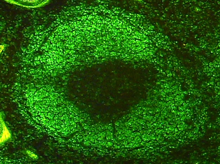
The immune response to vaccines and microbial pathogens is characterized by the spatial reorganization of leukocytes into microanatomical structures such as germinal centers and granulomas. Below we show a zoomed-in image of a lymph node section stained for B220, taken from C.Ji, D.Merl, T.Kepler, and M.West. Spatial mixture modelling for unobserved point processes: examples in immunofluorescence histology. Bayesian Analysis, 4(2):297--316, 2009.
Papers and presentations
- I. Manolopoulou, T. Kepler and D. Merl (2012) Mixtures of Gaussian Wells: Theory, Computation and Application in Immunofluorescence Histology. Computational Statistics and Data Analysis.
- Summer School in Computational Immunology, Santa Fe Institute (Aug 09) "Analysis of Spatial Data and Tissues".
- I. Manolopoulou, X. Wang, C. Ji, H. Lynch, S. Stewart, G. Sempowski, S. Munir Alam, M. West, T. Kepler, 'Statistical Analysis of Cellular Aggregates in Immunofluorescence Histology' Duke discussion paper (2009).