Close
Close

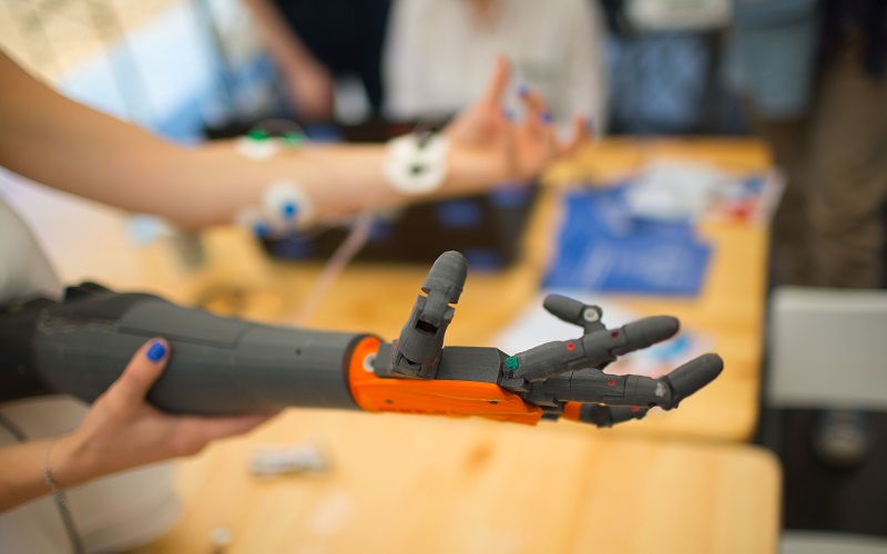
3D Models of Health and Disease
Our work
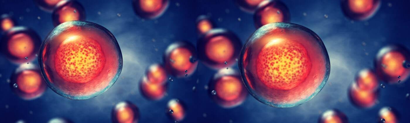
The Centre for 3D Models of Health and Disease
Principal Investigators


Post-doctoral fellows
Dr Deniz Bakkalci

PhD students


Anuja Upadhyay


Dr Dechao Feng


Lisa Purk


Kyle Saunders

Alumni
- Dr Alvena Kureshi
- Dr Rawiya Al Hosni
- Dr Judith Pape
- Dr Philip Barrett
- Dr Auxtine Micalet
Select publications
- Bakkalci D, Al-Badri G, Yang W, Nam A, Liang Y, Khurram SA, Heavey S, Fedele S, Cheema U (2023). Spatial transcriptomic interrogation of the tumour-stroma boundary in a 3D engineered model of ameloblastoma. Mater Today Bio. 2023 Dec 21;24:100923.
- Gupta P, Bermejo-Rodriguez C, Kocher H, Pérez-Mancera PA, Velliou EG (2024). Chemotherapy Assessment in Advanced Multicellular 3D Models of Pancreatic Cancer: Unravelling the Importance of Spatiotemporal Mimicry of the Tumor Microenvironment. Adv Biol (Weinh). 2024 Feb 7:e2300580.
- Bakkalci D, Al-Badri G, Yang W ... Cheema U (2023). Engineering a metastatic stroma directs the osteosarcoma tumour transcriptome in a spatially specific manner. Applied Materials Today, Vol. 35, 2023, 101994.
- Purk L, Kitsiou M, Ioannou C, El Kadri H ... Velliou EG (2023). Unravelling the impact of fat content on the microbial dynamics and spatial distribution of foodborne bacteria in tri-phasic viscoelastic 3D models. Sci Rep. 2023 Dec 9;13(1):21811.
- Micalet A, Tappouni LJ, Peszko K, Karagianni D, Lam A ... Cheema U (2023). Urokinase-type plasminogen activator (uPA) regulates invasion and matrix remodelling in colorectal cancer. Matrix Biol Plus. 2023 Nov 15;19-20:100137.
- Gupta P, Pérez-Mancera PA, Kocher H, Nisbet A, Schettino G, Velliou EG (2020). A Novel Scaffold-Based Hybrid Multicellular Model for Pancreatic Ductal Adenocarcinoma-Toward a Better Mimicry of the in vivo Tumor Microenvironment. Front Bioeng Biotechnol. 2020 Apr 24;8:290.
- Shepherd DW, Norris JM, Simpson BS, Player DJ, Whitaker HC (2022). Effects of photobiomodulation therapy on regulation of myogenic regulatory factor mRNA expression in vivo: A systematic review. J Biophotonics. 2022 Feb;15(2):e202100219.
- Pedersen K, Vanhoestenberghe A & Heavey S (2022). Urgent action is required to increase sustainability in in vitro modelling. In vitro models.
- Barrett P, Quick TJ, Mudera V & Player DJ (2022). Neuregulin 1 Drives Morphological and Phenotypical Changes in C2C12 Myotubes: Towards De Novo Formation of Intrafusal Fibres In Vitro. Frontiers in Cell and Developmental Biology, 9, 760260.
- Grillo A, Hyder Z, Mudera V & Kureshi A (2022). In vitro characterisation of low-cost synthetic meshes intended for hernia repair in the UK. Hernia, 2022 Feb;26(1):325-334. Epub 2021 Apr 2.
- Mudera V, Whitehead-Clarke T, Kureshi A, et al (2021). The experimental methodology and comparators used for in vivo hernia mesh testing: a 10-year scoping review. International journal of science and research methodology. Hernia.
- Ravi B, Kapoor M & Player DJ (2020). Feasibility and reliability of a web-based smartphone application for joint position measurement. Journal of Rehabilitation Medicine, 53(5), 1–6.
- Barrett P, Quick TJ, Mudera V & Player DJ (2020). Generating intrafusal skeletal muscle fibres in vitro: Current state of the art and future challenges. J Tissue Eng. 2020 Dec 29;11:2041731420985205.
- Wishart G, Gupta P, Schettino G, Nisbet A, Velliou E (2021). 3D tissue models as tools for radiotherapy screening for pancreatic cancer. Br J Radiol. 2021 Apr 1;94(1120):20201397.
- Mudera V, Player DJ & Kureshi A (2019). Engineering of Collagen as a Functional Biomaterial. Comprehensive Biotechnology, Third Edition. Elsevier.
Related courses