Molecular Imaging across UCLH BRC Using Radionuclide Hybrid Imaging Techniques
Our purpose is to develop Molecular Imaging that translates the world class biology being performed on the UCL campus to explicitly address the challenge of overcoming the major diseases inflicting our patients at UCLH.
The Institute of Nuclear Medicine is the largest single UK department committed to radioactive tracer methodology and has introduced to the UK several technologies and techniques in to humans (e.g. PET/CT, use of Rubidium-82 and gallium-68 generators, solid state cardiac-specific SPECT). It has an international reputation to support a range of patient driven research activity across disciplines and this capability extends to the support of other key development platforms. The Institute of Nuclear Medicine has had a consistently high research output amongst Nuclear Medicine departments internationally. We have taken delivery of the 1st PET/MRI scanner in the UK (6th in the world), which allows us, for the first time, simultaneous acquisition of data from two imaging modalities. This will complement our existing 2 PET/CT and 5 SPECT scanners. The Institute is in a unique position in the UK to deliver an experimental medicine frontier imaging programme across the UCL campus. We deliver early phase human studies using PET and SPECT ligands and imaging, trying to measure biological signals that help us understand pathogenesis and phenotype disease, and to risk stratify our patients.
Last year we introduced 5 new PET tracers imaging on campus in order to perform 1st in man studies.
In this Experimental Medicine Programme, we will focus on 1st in man studies, improving diagnostics, patient stratification and response assessments. We will perform in vivo studies on patients using dual signal imaging techniques to investigate across diseases the complex relationships between angiogenesis, perfusion, hypoxia and apoptosis and how these processes are modified by various cell receptors e.g. αvβ6 integrin. We will use more specific ligands brain diseases e.g. amyloid deposition.
The importance of mechanistic imaging is becoming increasingly recognised. Our dual signal imaging techniques allow two methodologies to be cross validated. In addition, dual source imaging affords the ability to acquire complementary imaging data. The latter may be utilised to tease out the pathogenesis of disease and may yield important insights into the targeting of treatments. For example in cancer, it can identify two pathways for therapeutic intervention thus reducing the opportunity for tumour resistance. We will use PET/MRI, PET/CT and SPECT/CT hybrid imaging techniques in combination with novel radionuclides and MRI/CT ligands and techniques across a wide spectrum of patients. We validate our work using surgical samples accurately co-registered to our images allowing us to compare our imaging signals with genomic and proteomic tissue signatures. We continue our active collaborations and seek further interactions across UCL basic scientists (and CRICK in the future) in pursuit of novel biology derived from histological samples to compare with our novel imaging parameters.
Research Output
In the last 3 years we have published 18 times in the Journal of Nuclear Medicine (ranked 1st out of 113 Imaging Journals), by far the greatest peer reviewed output of any other similar UK Institution.
Current Collaborations
We have extensive research interactions with a broad range of UCL biomedical scientists including, Queen Square (neurovascular, epilepsy, neurodegeneration, neuroinflammation), Cardiovascular Institute (infarction and cardiac muscle diseases), Centre of Respiratory disease (inflammation and lung cancer), Cancer Institute and Surgery (Upper GI, Lower GI and Vascular).We have collaborations with other UCL CBRCs (GOSH- Congenital heart disease, early atheroma, cancer) and UCLP sites -RFH, Barnet (cancer and cardio/neurovascular) and further afield. N.E Herts NHS Trust (fibrosis and cancer). We collaborate with other CBRCs- Imperial (cancer and Inflammation), Cambridge (Inflammation, cancer and vascular) and Royal Marsden (Cancer) and other CCIC/ECMCs -KCL, Imperial and Royal Marsden).
Industrial collaborations
We have active collaborations with GE Healthcare, Siemens, Spectrum Dynamics, Advanced Accelerator Applications, COVIDIEN and GSK. We are in various stages of negotiating further contracts related to use of novel tracers with GE (Angiogenesis, Inflammation and dementia tracers), SIEMENS (Hypoxia and dementia tracers) and GSK (Lung fibrosis and now moving into other organs) with a new interaction with TEX RAD (Inflammation, Cancer, cardiovascular). New interactions are occurring with Roche (inflammation and cardiovascular) and Oncovision (cancer).
Medical Physics
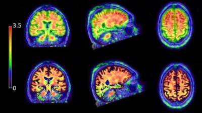
An Alzheimers patient imaged with Flutemetamol (GE). The lower Images are higher resolution than the upper row due to novel image corrections. This is vital for signal quantification in order to detect early disease.
The clinical research programmes are underpinned by innovative development in Medical Physics, which focuses on quantitative studies in emission tomography (SPECT and PET) and multimodal imaging. The group has a reputation in development of SPECT instrumentation and in image reconstruction for PET and SPECT with projects funded through EPSRC, CRUK and EC and industrial collaborations. The group develops algorithms for improved quantification correction for factors such as attenuation, scatter, resolution and motion. Much of this development exploits the information available from dual modality studies. There is a close collaboration with researchers at the Centre for Medical Image Computing (CMIC at UCL). Active development work includes: cardiac studies (validation of partial volume correction in conjunction with Yale University, improved attenuation correction for application in study of angiogenesis in patients treated with stem cells; algorithms to facilitate dual radionuclide studies using novel SPECT detectors); inflammatory diseases (corrections for tissue fraction so as to provide more sensitive biomarkers in assessing treatment of pulmonary fibrosis); neurological studies (development of partial volume correction techniques applied to early detection of dementia); cancer (optimised design of SPECT systems specifically for use in study of cancer. The group works closely with physicists in Radiology and CMIC to exploit the information from the new PET/MRI.
The clinical research programmes are underpinned by innovative development in Medical Physics, which focuses on quantitative studies in emission tomography (SPECT and PET) and multimodal imaging. The group has a reputation in development of SPECT instrumentation and in image reconstruction for PET and SPECT with projects funded through EPSRC, CRUK and EC and industrial collaborations. The group develops algorithms for improved quantification correction for factors such as attenuation, scatter, resolution and motion. Much of this development exploits the information available from dual modality studies. There is a close collaboration with researchers at the Centre for Medical Image Computing (CMIC at UCL). Active development work includes: cardiac studies (validation of partial volume correction in conjunction with Yale University, improved attenuation correction for application in study of angiogenesis in patients treated with stem cells; algorithms to facilitate dual radionuclide studies using novel SPECT detectors); inflammatory diseases (corrections for tissue fraction so as to provide more sensitive biomarkers in assessing treatment of pulmonary fibrosis); neurological studies (development of partial volume correction techniques applied to early detection of dementia); cancer (optimised design of SPECT systems specifically for use in study of cancer. The group works closely with physicists in Radiology and CMIC to exploit the information from the new PET/MRI.
Radiochemistry
Our Radiochemistry lab was a new CBRC funded initiative established in 2008 as part of collaboration between UCL Chemistry, Division of Medicine and UCLH. We have constructed state-of-the-art facilities for radiolabelling, tracer development and pre-clinical imaging studies, which underpins experimental imaging studies in man. The investments in radiochemistry at UCL/UCLH has been leveraged by the formation of the Pan London Cyclotron unit (Imanova), which will enable access to new 18F-labelled tracers for experimental human studies at INM, as well as PET imaging with carbon-11 tracers at Hammersmith hospital. Radiochemistry plays an integral role in nuclear imaging, and will be instrumental to (i) enable access to a wider range of PET tracers to support the experimental medicine programme, (ii) provide blood metabolite analysis for absolute quantification of PET imaging data, (iii) accelerate translation from pre-clinical studies to experimental imaging studies in man, and (iv) development of new radiotracers for human studies.
In Oncology, the Institute of Nuclear Medicine pioneered a number of imaging techniques in man, including sentinel lymph node scintigraphy, somatostatin receptor imaging in neuroendocrine tumours and metabolic flow imaging combining PET with CT and MRI in various cancers. In the last 12 months four PET oncology tracers have been introduced on campus for study of hypoxia (FMISO), angiogenesis (Fluciclatide), Dopamine transporters (F-DOPA), and membrane metabolism (18F choline). Collaborating with industry we have just employed a software engineer to assess tumour heterogeneity with imaging.

|
Assessing Tumour Phenotype and Heterogeneity
In cancer we apply a Stratified Experimental Medicine approach. A priority of our programme is to use imaging to access tumour heterogeneity and phenotype tumours for targeted therapy.

|
We build on our last 5 years work with the Imaging group at UCH, performing in vivo human studies using dual signal imaging techniques to investigate the complex relationships between tumour vascularity, hypoxia and glucose metabolism, and now are investigating how membrane receptors can modify these processes e.g. somatostatin, αvβ6 integrin and TSPO.

|
|
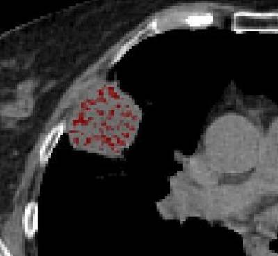
By using surgical patients we compare our imaging signals with tumour genomic and proteomic tissue signatures aided by accurate image/ histology co-registration techniques using ex-vivo CT. We now collaborate with UCL and Barts biologist using on novel markers (e.g. hormonally upregulated neu-associated kinase) to correlate with imaging.
Novel and Targeted Therapeutics
Collaboration is intensifying between the Institute of Cancer's ECMC Group, UCH CRF and Nuclear Medicine, with studies of novel agents and antibodies. Targeted tumour antibodies are being pioneered using the new Cancer Institute's SPECT/CT that is located within our department. This equipment is also being used to assess a first in man study of a liposomal drug delivery system of a novel antimitogenic in collaboration between a US company (Eisai) and Cancer and Nuclear Medicine Institutes.
In 2011 with GOSH, we carried out a first in man study using therapy with Lu-177-DOTATATE in children with Neuroblastoma. Also with GOSH and with Royal Marsden we are performing the first use of the novel PET ligand, 124I-MIBG PET in neuroblastoma funded by CRUK (DDO). With industry, Barts and Imanova, we are to perform a 1st in man cancer study with a αvβ6 PET ligand; (TGF beta pathway) and we investigate its effect on tumour angiogenesis, hypoxia and apoptosis. The ligand is an ideal target for therapeutic intervention for the Cancer Institute's antibody group.
With oncologists and biologist at UCL and Barts/ QMUL, we are performing NIHR and industrial studies (GE/Pfizer), using sequential PET techniques to predict response with novel therapies renal cell cancer.
We are offering key support to UCL/H prostate cancers studies, aiding successful grants, combining novel PET ligands with MRI techniques. A Prostate Action Group Grant is now in application.
With Royal Free surgeons and oncologists and with Oncovision we are combining PET Mammography (1st in UK) with or new PET/MRI scanner (1st in UK) to guide personalise breast cancer therapy. The new PET MRI scan has resulted in multiple new cancer proposals.
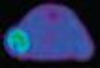
Figure of a PET scan from a pancreatic carcinoma mouse model demonstrating the first in vivo imaging of αvβ6 expression (green colour). In Q1 2013 with industry, Barts and Imanova, a first in man normal volunteer will be performed using this novel tracer. This will be followed by a first human cancer study.
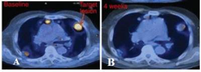
A patient with pulmonary metastatic renal cell cancer. At baseline PET (A), the metastases are metabolically active (yellow colour). Following novel targeted treatment (B) there has been a dramatic metabolic response.
Stem Cells
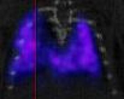
Stem cell tracking in a rat at CABI and with the Centre of Respiratory Research.
The SPECT image shows signal accumulation from implanted stem cells accumulating in the lungs. This study is translating into a first in man study which is being applied to the delivery of therapy to lung cancer.
Having introduced Rb myocardial perfusion PET/CT to Europe in 2006, we then pioneered dual imaging techniques to investigate both cardiac and arterial disease. We have close links with multiple cardiovascular groups across UCL including CBRCs. We aim to use and SPECT ligands in combination with CT and MRI to investigate to address the challenges identified in the BRC programme.
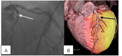 Introducing 82-Rb into Europe, allowed us to develop cardiac hybrid PET CT imaging. Image A- an invasive angiogram shows LAD stenosis (white arrow). PET confirms LAD ischaemia. The fused cardiac PET (B) (colour scale) and CT angiogram image confirms a stenosed LAD (black arrow) and anteroseptal ischaemia (orange) in the left ventricle. Good perfusion (yellow) elsewhere. These techniques will enhance our use of PET/MRI. |
Evaluation/Monitoring Cardiovascular Risk in the Young and Asymptomatic
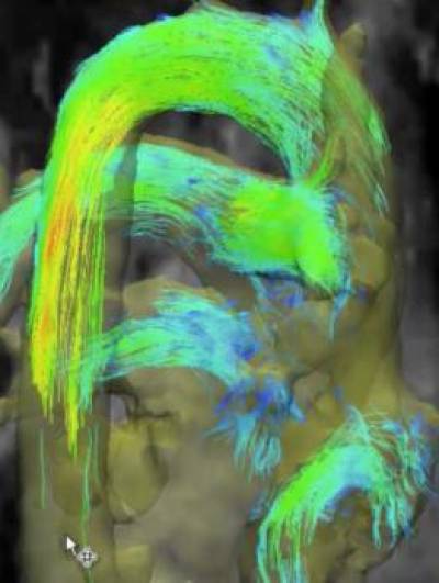 4D MRI techniques developed at GOSH allowing rapid acquisition and showing blood flow on the aorta. | The advent of PET/MRI has opened up new opportunities for cardiovascular risk detection and therapeutic monitoring, especially in the young with implications for mass screening for early disease detection. Combining sensitive PET detectors with MRI rather than CT, reduces radiation exposure allowing use in the young, asymptomatic and repeated examinations needed for treatment monitoring. Collaborating with GOSH and Industry, we are specifically addressing early atheroma formation in the young. Applying GOSH techniques to PET/MR we will identify aortic blood flow and measure shear stresses on the aorta, whilst simultaneously using PET ligands to measure angiogenesis at these sites to investigate mechanisms and detection of early atheroma formation. We have been performing aortic aneurysm research in asymptomatic patients. Working with Vascular Surgeons at UCH, with UCL P hospitals and in collaboration with University of Sussex, we have shown we can use FDG PET to predict future aortic aneurysm growth. We are now correlating PET imaging biomarkers with novel serum proteomics with the Centre of Clinical Pharmacology. |

|
Monitoring Response of CRP inhibiters & Novel Cardiovascular-protective Interventions
We are internationally recognised for our vascular PET imaging protocols that reduced imaging times by a third, paving the way for therapeutic monitoring of novel agents e.g. CRP inhibitors. Multiple drug companies invite us to monitor therapy. We are now to collaborating with the Centre for Cardiovascular Genetics, performing a novel cytokine intervention study using PET as an imaging surrogate.
Pathophysiological Determinants of Beneficial Response to Surgery
Continuing the investigation of the Lancet published research by the Hatter examining preconditioning in CABG, we will use PET/MRI to investigate the mechanism of the conditioning. We are working with Siemens and the Hatter using apoptosis and angiogenesis PET ligands in NSTEMI patients undergoing ischaemic preconditioning.
Collaboration with Yale
Having performed a first in man study of a solid state PECT detector for myocardial perfusion in with US centres (published in JACC), we apply this imaging to cardiac stem cell research with Yale. We are investigating angiogenesis in ventricular remodelling using a SPECT ligand in stem cell therapy.
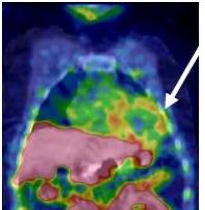 | In collaboration with Yale, GE/ Barts and Centre of Cardiovascular Biology, we are investigating the role of angiogenesis in ventricular remodelling after the administration of stem cells. The SPECT/CT image shows the biodistribution and myocardial uptake (arrow) of the angiogenesis ligand (RGD peptide- αvβ3 integrin). |
Complimentary Imaging to CMR in Cardiac Amyloid, Fibrosis and Inflammation
We will complement the work being performed on myocardial fibrosis at the Heart hospital on CMR by using a fibrosis mediated PET ligand (αvβ6 integrin). Also in collaboration with the heart Hospital as well as the Centre for Cardiology in the Young, we are building on the use of our hybrid imaging using Rubidium-82 myocardial PET and cardiac CT angiography in patients with hypertrophic cardiomyopathy.
In collaboration between the Centre for Respiratory Diseases and the Heart Hospital, we are using PET ligands that are involved in the mediation of inflammation to investigate patients with myocardial sarcoid and differentiating these individuals from those with arrhythmogenic right ventricular dysplasia.
 Anterobasal Myocardial uptake of uptake of FDG confirmed on cardiac MRI (arrow). A potential area where the new PET MRI scanner can show added value. |
CT Textural Analysis in Cardiovascular Causes of Sudden Death
UCL cancer investigators have shown the importance of genomic heterogeneity in tumours just published in NEJM. INM have pioneered measurements of CT heterogeneity in cancer using the CT component of PET CT. We are now relating this to genomics and also investigating properties that could be applied to the cardiovascular system. We a have shown heterogeneous aneurysms confer a poor prognosis and will apply the technology to the myocardium in in inherited muscle disorders e.g. HCM.

First study measuring heterogeniety of aortic aneurysms using CT textural analysis. Our longitudinal study demonstrated that heterogenous aneurysms expanded more than homogenous aneurysms at 1 year.
The last 5 years has seen novel PET developments in these areas within our department. We have pioneered PET Imaging techniques in interstitial lung diseases that have established us as a leading International reputation in this field. We have shown that patients that had lung changes of typical idiopathic pulmonary fibrosis had highly metabolically active lungs on PET.
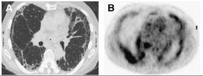 Idiopathic pulmonary fibrosis is a fibrotic lung disease with a median mortality of 3 years and no effective treatment. Above are images of a HRCT (A) and 18F-FDG PET (B) images from 74-y-old man with newly diagnosed Idiopathic pulmonary fibrosis HRCT image (A) shows typical pattern of usual interstitial pneumonia. Areas of most intense 18F-FDG uptake correspond to areas of parenchymal honeycombing on CT. These areas were traditionally believed to be burnt out end stage disease, but our research show that instead they represent hypermetabolic areas that should be amenable to pharmocological targeting. |
Our methodologies are being incorporated into drug discovery programmes. Indeed our work with GSK was chosen as an example of experimental medicine approach being translated directly into patient benefit. Using a novel GSK ligand we are performing the first human study using FDG PET as an early surrogate end point for monitoring therapy as well as using PET to titrate.
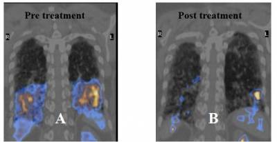 A patient with inflammatory lung disease (NSIP) secondary to a drug reaction. The pretreatment image (A) shows increase PET signal (Yellow/Blue) at the lung bases. This signal diminished following successful treatment. This proof of concept study has provided the rationale for successful grants with NIHR and GSK. |
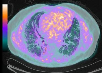
To further understand the mechanism and pathogenesis of idiopathic pulmonary fibrosis we are applying to the MRC's Experimental Challenge call with biologists and clinicians from the Centre of Respiratory disease and with collaborators from 2 other CBRCs (Royal Brompton and Cambridge). We are currently using different PET ligands, including tracers that can measure somatostatin receptors (collaboration with Covidien), angiogenesis (collaboration with GE) and hypoxia (collaboration with GSK).
|
Next year we are collaborating with Radiochemistry, GSK and the new Joint universities cyclotron facility (IMANOVA) to perform a first in man study of normal volunteers using a new PET integrin ligand that is believed to be key mediator of these diseases as well as in cancer. This study will then be followed by the first use of this ligand in patients with fibrotic lung disease. We are taking our PET techniques outside of the lung to image fibrosis in others organs (Liver and kidney) in combination with novel CT/MRI techniques using hybrid PET/CT and PET/MRI scanners.
In addition to the novel ligands above we are also in negotiations with local industry for a new fluorinated PET TSPO ligand. This mitochondrial receptor ligand is a key mediator for inflammation arising from many insults such as ischaemia, neurodegeneration, injury and stress. As such, the use of this receptor agonist will have implication for PET inflammation imaging in many diseases and can be employed in multiple systems. In the lungs it will complement our interstitial pulmonary disease programme. In the arterial system we will measure aneurysm expansion, in carotid atheroma we will correlate its uptake with endarterectomy specimens and in the myocardium we will use it as a novel method to image cardiac sarcoid. In the brain it will have a key role in imaging neuroinflammation.
In collaboration with the centre for respiratory diseases and Sussex University, we are translating work on cell trafficking performed at CABI and applying this to in vivo in humans. We are using the Indium labelled platelets and neutrophils to investigate possible cellular dysfunction in the lungs of patients with idiopathic pulmonary fibrosis and other forms of interstitial lung disease.
Finally we are applying our inflammation PET monitoring techniques to beyond the lungs. We are working with industry and the Centre for Cardiovascular Genetics which have been investigating the genetics of Interleukin biology. We will be providing frontier PET inflammation measurement techniques to monitor novel anti-inflammatory pharmacological intervention in the aorta.
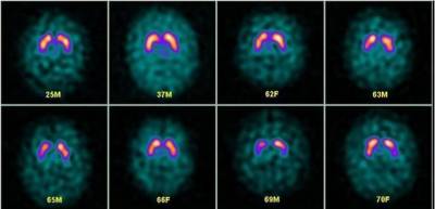
The INM pioneered several imaging techniques assessing patients with neurodegenerative disorders. A 1st in man study was performed with Tc-99m HMPAO, which became the 1st conventional radionuclide tracer to traverse the intact blood brain barrier, permitting blood flow imaging in the human brain. The INM had a number of firsts in this area, SPECT maps of the D2/D3, benzodiazepine, sigma-1 and 5HT2 receptors in the brain.
In direct pursuit of pre-symptomatic detection of disease, in 2012 we have introduced the first amyloid plaque PET agent to UCLH in collaboration with the Institute of Neurology.
Although, Nuclear Medicine has been part of the Body Imaging theme, it provides SPECT, PET and radioligand imagining for multiple campus neuroscience researchers. In total 17 separate UCL researchers use or intend use these technologies for research across a wide range of diseases, (epilepsy, psychiatry, multiple sclerosis, dementia, cognitive sciences and intracranial tumours).
Such is the extent of interaction, in collaboration with GE, Nuclear Medicine, Institute of Neurology and Chalfont are in exploration of setting up a satellite PET operation to facilitate early neuro-PET ligand development in human studies.
|
Pre-symptomatic Detection of Disease
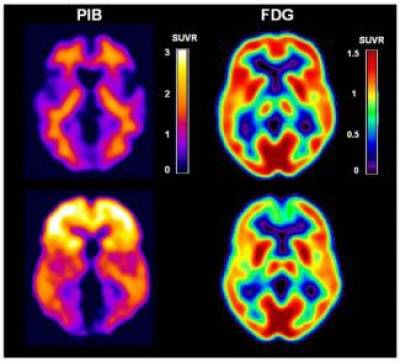
PET image "of 2 normal volunteers: one at low and one at high risk of AD. Right sided images show positive uptake in plaque, left images show glucose negative image in the plaque. The distribution of tracers on the lower row, indicates that individual is at high AD risk! The Upper row shoes a low risk patient(see 1st UCH images below.
In collaboration with the Dementia Research Centre and Neurodegenerative Diseases at the Institute of Neurology we have started a first trial of florbetapir (F-18 AV-45), an amyloid PET imaging agent, to investigate its role in focal dementia syndromes. The 1st UK study using amyloid plaque PET/MRI imaging is also being performed in collaboration with Sussex University.
INM is well placed to perform state-of-the-art image analysis on amyloid studies and other quantitative neurological studies, after the development of methods for improved partial volume correction applied to amyloid studies provided by GE Healthcare.
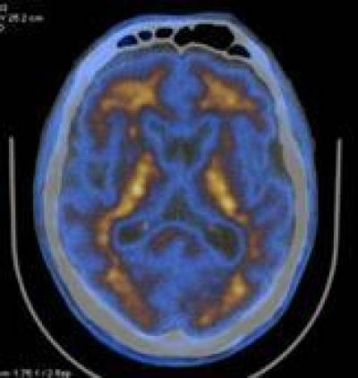 | Image from the 1st UCLH use of a beta amyloid PET ligand, showing low uptake of ligand (blue colour) in the grey matter thus the subject at low risk for Alzheimer's disease. |
Imaging beta amyloid and tau will become a major new area in the recently successful grant application to the Wolfson and the Experimental Neurology Centre. Multiplexing several radionuclide probes with MR sequences will offer an exciting new avenue for molecular imaging and phenotyping in vivo in man.
Stroke- Risk Stratification and Therapeutic Monitoring
Our multimodality imaging is enabling us to risk stratify patients for cerebral vascular accident. Moreover the imaging can provides quantitative imaging that then can be used to monitor risk reducing therapies. Our strength in quantifying these imaging signals has led to an international reputation for monitoring novel interventions.
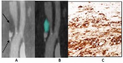
In collaboration with the Department of Brain Repair & Rehabilitation, the Stroke Research Group, Hyperacute Stroke unit and Vascular Surgery, we have developed multimodality imaging of carotid plaque with histological validation from resected specimens. Our data is advancing a pan UCL database, which will cross reference proteomic and genomic data. We are furthering this work now using Somatostatin receptor, hypoxia and inflammation (TSPO) imaging.
|
Psychiatry
In collaboration with the Mental Health Sciences Unit and GE we are using PET/MRI with the novel
amyloid PET ligand flutemetamol. We are investigating the role of amyloid deposition load in parkinsonian movement disorders and Lewy Body dementia.
Imaging to Guide Neurosurgical Intervention
With the Department of Clinical and Experimental Epilepsy we are using our new PET/MRI camera to investigate epilepsy. We are exploiting the PET and MRI simultaneous signal acquisition to temporally and spatially match cerebral metabolism with functional MRI.
Diagnostics and Therapeutic monitoring
We have just obtained an ECFP7 grant to develop a first in man SPECT MRI system. This is for imaging the brain and because it is SPECT it will increase the novel ligands we can use for both diagnostic and therapeutic purposes
Recently in collaboration with the Department of Motor Neuroscience & Movement Disorders at the Institute of Neurology we started a Phase II, randomised controlled trial evaluating.
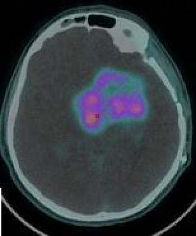 | In collaboration with GOSH, we are performing the first paediatric study using the novel PET tracer 18F Choline PET a biomarker of cell membrane metabolism, in the management of children with glioblastoma. The focus of colour fused on the black and white CT image of the brain depicts the region of metabolically active glioma as detected with PET. This work is being translated with major implication for urgently needed drug development. Other intracranial oncology research includes a 1st in man study of somatostatin analogue PET ligand with PET/MRI for treatment planning in meningioma. |
 Close
Close

